Method of colorectal cancer detection by using radiolabeled anti-GRP78 peptide

a colorectal cancer and radiolabeled technology, applied in the field of colon cancer detection, can solve the problems of increasing the cost of treatment, increasing the risk of infection, and taking a long time to detect antibodies,

Inactive Publication Date: 2014-04-08
INST NUCLEAR ENERGY RES ROCAEC
View PDF1 Cites 4 Cited by
  • Summary
  • Abstract
  • Description
  • Claims
  • Application Information

AI Technical Summary

Benefits of technology

[0006]The main purpose of the present disclosure is to detect position and distribution of colorectal cancer tumor through nuclear imaging, or to identify stage of colorectal cancer by shading value in tumor, where the present disclosure is thus a safe and noninvasive clinical method for diagnosing and tracing level and distribution of colorectal cancer before and after treatment.

Problems solved by technology

In addition, detection through colonscopy takes time and patient may feel uncomfortable, even in danger of enterobrosis.
However, because antibody may cause immune response, antibody may take a long time to reach position of tumor through the circulatory system and the patient may have to be exposed to radio nuclide for a long time too.
Furthermore, the expense may be thus increased.
Hence, the prior arts do not fulfill all users' requests on actual use.

Method used


Image

  • Method of colorectal cancer detection by using radiolabeled anti-GRP78 peptide
  • Method of colorectal cancer detection by using radiolabeled anti-GRP78 peptide
  • Method of colorectal cancer detection by using radiolabeled anti-GRP78 peptide

Examples


Embodiment Construction

[0015]The following description of the preferred embodiment is provided to understand the features and the structures of the present disclosure.

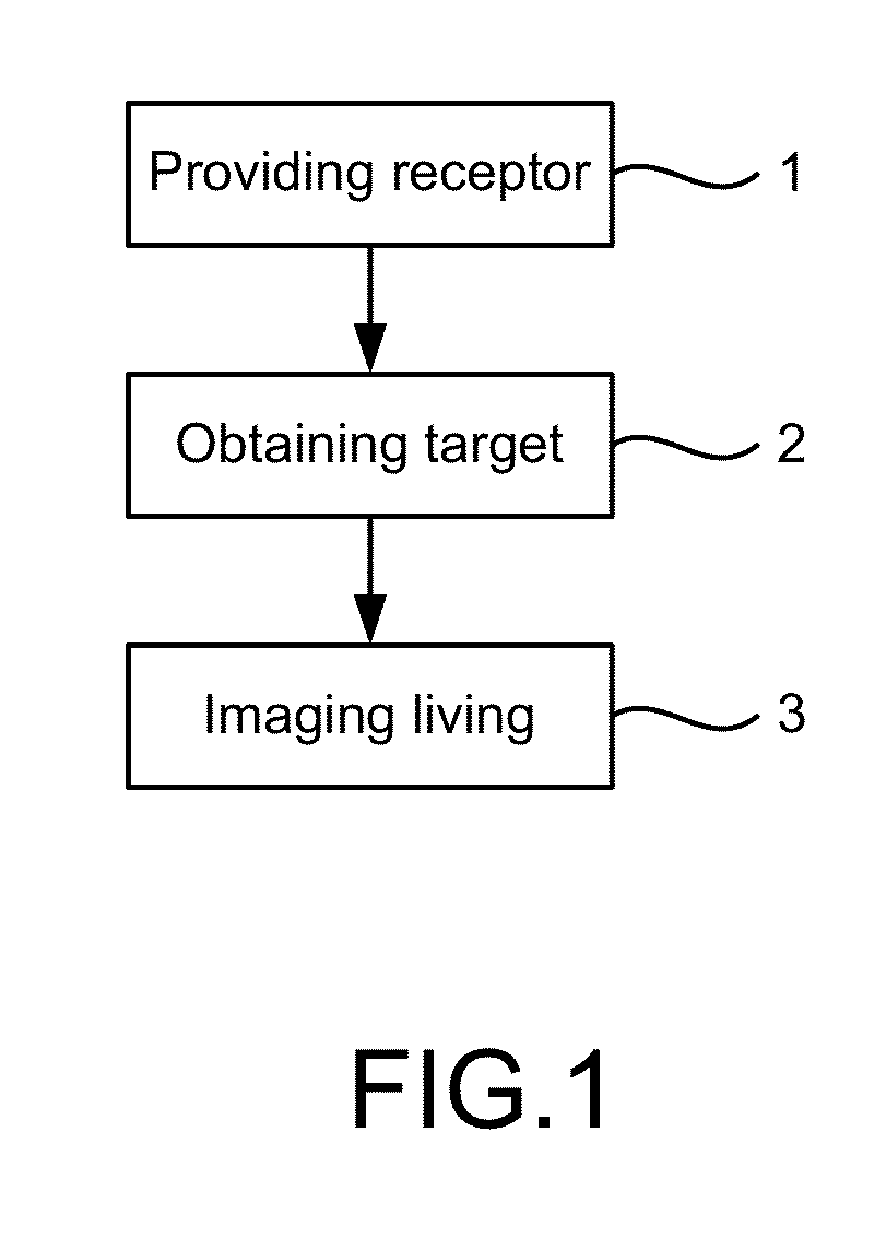
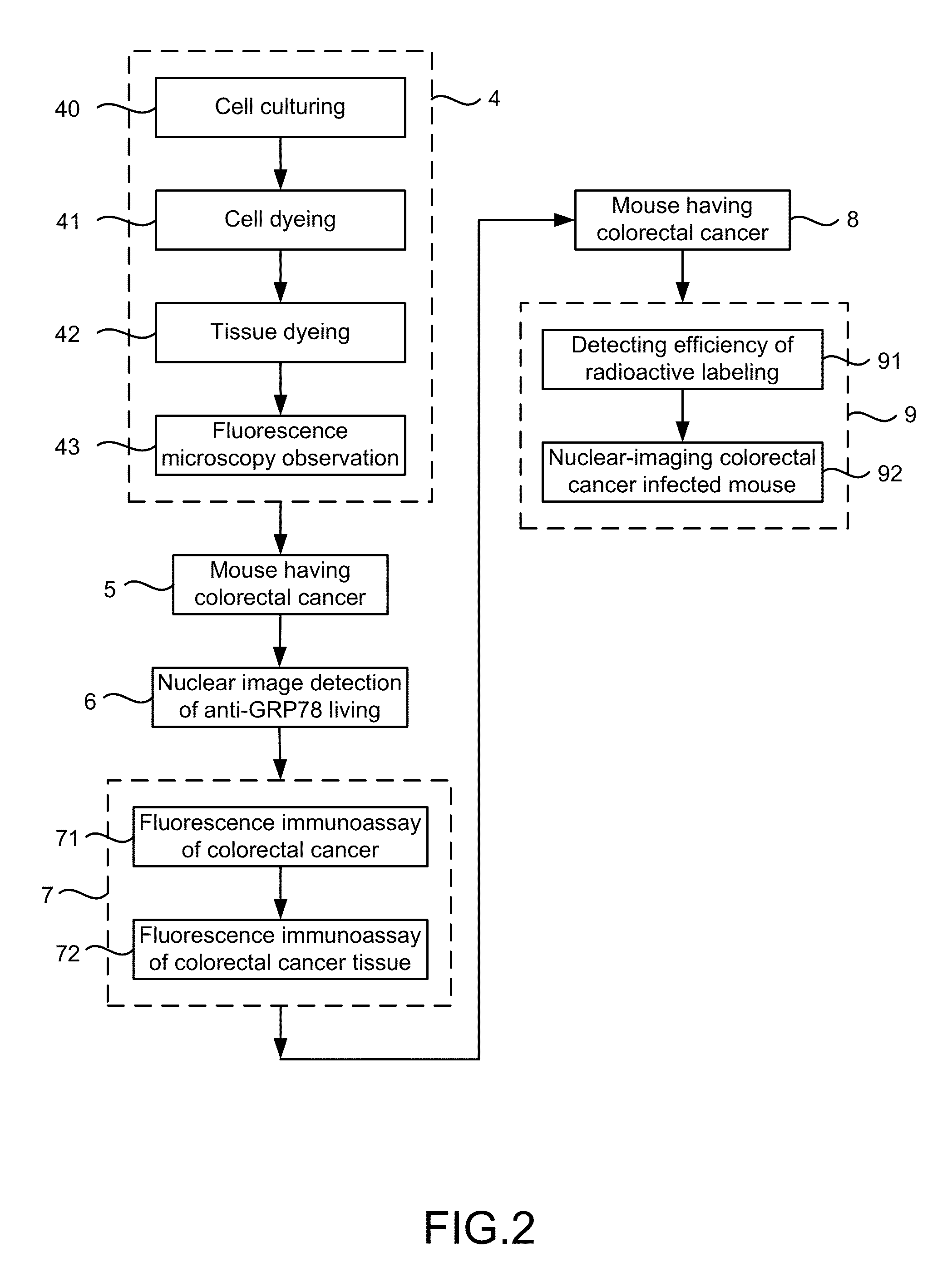
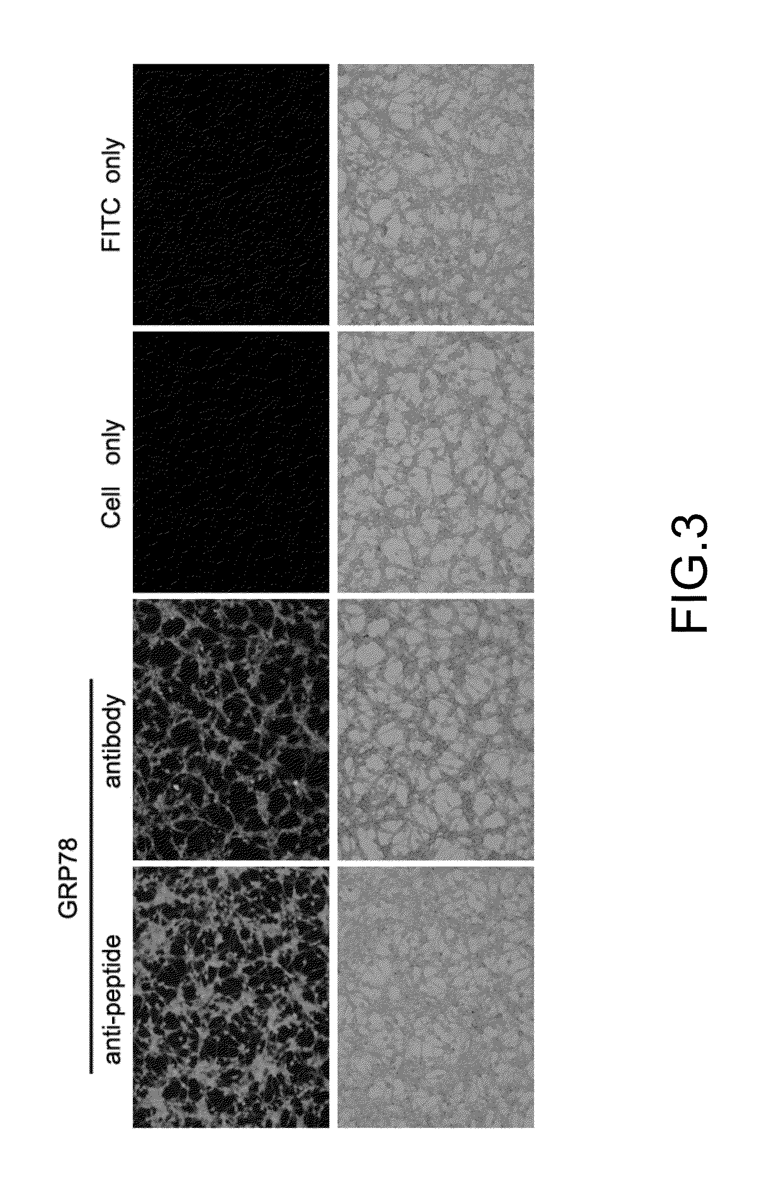
[0016]FIG. 1 to FIG. 6 are a flow view showing a preferred embodiment according to the present disclosure; a flow view showing confirmation of the preferred embodiment; a view showing immunofluorescence cell staining of colorectal cancer; a view showing immunofluorescence tissue staining of colorectal cancer; a view showing labeling effects after 1 hour and 1.5 hour; and a the view showing microSPECT / CT. As shown in the figures, the present disclosure is a method of colorectal cancer detection by using radiolabeled anti-GRP78 peptide, where detection is noninvasive on taking a sample from living for diagnosing and tracing colorectal cancer before and after a treatment. The method according to the present disclosure comprises the following steps:

[0017](a) Providing receptor 1: An anti-peptide receptor of glucose regulated protein 78 (GRP78) o...


PUM

PropertyMeasurementUnit
thicknessaaaaaaaaaa
wavelengthaaaaaaaaaa
computed tomographyaaaaaaaaaa

Abstract

The present disclosure describes techniques used for colorectal cancer detection. Position and distribution of colorectal cancer tumor are detected through nuclear imaging. Alternatively, stage of colorectal cancer is identified by shading value in tumor. Thus, the present disclosure provides a safe and noninvasive clinical method for diagnosing and tracing level and distribution of colorectal cancer before and after treatment.

Description

TECHNICAL FIELD OF THE DISCLOSURE[0001]The present disclosure relates to colorectal cancer detection; more particularly, relates to detecting position and distribution of colorectal cancer tumor through nuclear imaging, or identifying stage of colorectal cancer by shading value in tumor, where the present disclosure is thus a safe and noninvasive clinical method for diagnosing and tracing level and distribution of colorectal cancer before and after treatment.DESCRIPTION OF THE RELATED ARTS[0002]Colorectal cancer is one of the most common cancers. Even some said it is the secondary major cause to death in the world.[0003]Recently, happening ratio of colorectal cancer is rising. Colorectal cancer in the early stage has a five-year survival rate of more than 90% if treatment is applied before expansion of the cancer cells. But, only ⅓ of the cancer patients are found before cancer metastasis. If metastasis to lymph happens, the five-year survival rate will be reduced to about 50%. Once...

Claims


Application Information

Patent Timeline
Patent Type & Authority Patents(United States)
IPC IPC(8): A61K49/00C07K7/06A61K38/08A61K49/04A61K49/14
CPCA61K51/08A61K38/08A61K49/0004A61K49/14C07K7/06
Inventor GUAN, SIAO-SYUNCHENG, CHUN-CHIALEE, SHUI-CHENGWU, HSIEN-MING
Owner INST NUCLEAR ENERGY RES ROCAEC
Why Eureka
Try Eureka