
Method for directed cell in-growth and controlled tissue regeneration in spinal surgery

a spinal surgery and directed cell technology, applied in the direction of biocide, prosthesis, drug composition, etc., can solve the problems of high pain, high percentage of perineural fibrosis and adhesion, need for additional surgery, etc., to prevent and minimize uncontrolled adhesion and scar formation

Active Publication Date: 2014-04-22
BAXTER INT INC +1
View PDF174 Cites 0 Cited by
  • Summary
  • Abstract
  • Description
  • Claims
  • Application Information

AI Technical Summary

Benefits of technology

The patent describes a new type of collagen-based material that can be used as a therapeutic implant. Unlike other collagen-based materials that are viewed as foreign and encapsulated, this material acts as a membrane that guides the growth of cells and helps to regenerate tissues. It also prevents unwanted cell growth and scar formation, which can lead to adhesion and fibrosis. This material is biodegradable and can be used to promote the healing of spinal structures. It is toxic and fluid-tight, which prevents the distribution of blood and necrotic material that can lead to adhesion formation. Overall, this material has the potential to improve the healing process and minimize the formation of scar tissues.

Problems solved by technology

Internal scarring, peridural and perineural fibrosis and adhesions after spinal surgery are well known and undesired side effects of said surgery.
These conditions lead to a high percentage to pain, motion difficulties and often to the need for additional surgery.
In these examples, a gel is distributed in an uncontrolled manner in the application area and once applied, the gel distribution cannot be easily manipulated or corrected.
Further, these anti-adhesion gels have limited success as a barrier and have undefined layer thickness.
These anti-adhesion gels have low hemostatic properties, if any at all, and provide no wound healing support functions and do not direct cell growth and tissue regeneration.
The DURAGEN PLUS barrier, which is of bovine origin, is not very shape stable, which means that its shape and position is difficult to correct after application.
Further, the DURAGEN PLUS barrier does not have a high tensile strength and elasticity.
And due to the fact that the DURAGEN PLUS barrier is porous it is consequently not fluid-tight (impermeable to fluids) and therefore has limited success as a barrier function.

Method used


Image

  • Method for directed cell in-growth and controlled tissue regeneration in spinal surgery
  • Method for directed cell in-growth and controlled tissue regeneration in spinal surgery
  • Method for directed cell in-growth and controlled tissue regeneration in spinal surgery

Examples


examples

Adhesion and Fibrosis Formation, Basics and Study Goal

[0067]Excessive scar tissue formation and adhesion is a serious problem in spine surgery, frequently causing radicular pain and physical impairment, e.g., failed lumbar disc surgery or post discectomy syndrome. The incidence of dura mater lesion in case of revision surgery is reported between 5 and 10% in the literature.

[0068]The most important critical parameters for the postoperative development and extent of adhesion formation are:[0069]the preoperative situation of the surgical area (existing adhesions / fibrosis, inclusive a genetically predisposition of the patient);[0070]the intraoperative performance of the surgery (extent of wound area, avoidance of injuries of tissues and certain anatomical structures (e.g. membranes), carefulness of hemostasis, avoidance of drying out of tissues etc.); and,[0071]intraoperative measures for prevention and minimization of postoperative adhesions, e.g. implantation of anti-adhesive implants...

example i

Materials and Methods: Animal Experiments Showing Reduced Adhesions in Spinal Surgery Using the Biofunctional Collagen Foil Biomatrix

A) Materials and Methods as shown in FIGS. 7-9:

Animals: New Zealand White rabbit, both genders, weight 4 kg. Origin: Charles River, Sulzfeld, Deutschland or Harlan Winkelmann, D-33178 Borchen.

Models and Groups:

Use of a biofunctional collagen foil biomatrix: Laminectomy, exposure of the spinal nerve root and application of the collagen foil biomatrix. Each group includes 20 animals.

Group 1: Laminectomy, Skin closure (Control).

Group 2: Laminectomy, Covering with collagen foil biomatrix.

Surgery:

[0075]The animals were positioned in a prone position and secured with the surgical area (lumbar spine) shaved. Under sterile conditions, after median incision of the skin, the Para vertebral musculature was detached from the spinous processes and the laminae of the lumbar vertebral spinal column exposed. Hemilaminectomy of the 3rd and 4th lumbar vertebra was condu...

example ii

Collagen Foil Biomatrix One Week After Implantation in Human Patient

[0077]The human patient (age: 18, gender: female) was operated in the interval of 7 days in the frame of a routine surgical treatment (epilepsy). The collagen foil biomatrix was implanted epidural during the first surgery (prevention of CSF leaks). During the second surgery, the collagen foil biomatrix implant was removed routinely before the reopening of the dura. After one week of epidural implantation, the collagen foil biomatrix is still mechanically stable and removable as shown in FIG. 10.

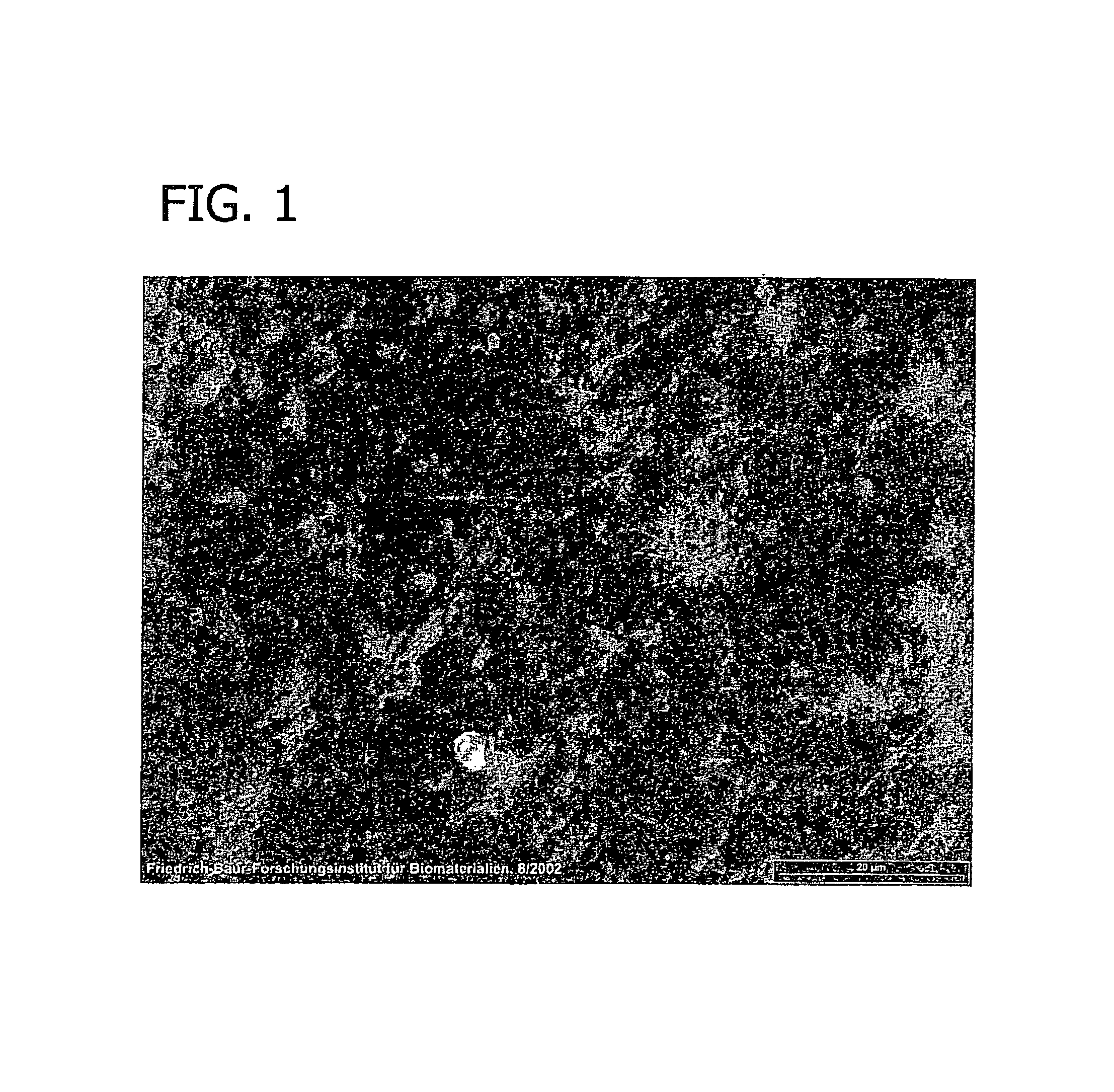
[0078]FIG. 11 is a sectional view of the collagen foil biomatrix of FIG. 10 one week after implantation. As can be seen, fibroblasts are growing into the collagen foil biomatrix, directed by the multi-layer structure. The penetration in longitudinal direction is about 220 to 320 μm.

[0079]The speed of the directed in-growth of repair cells along the multi-layered structure is about 10 to 15 times higher in longitudinal directi...


PUM

PropertyMeasurementUnit
thicknessaaaaaaaaaa
thicknessaaaaaaaaaa
thicknessaaaaaaaaaa

Abstract

The present invention relates to a method for directed cell in-growth and controlled tissue regeneration to prevent post-surgical or post-traumatic adhesion and fibrosis formation on the injured surface of a tissue selected from the group consisting of spinal column tissue, dura mater, and spinal nerves in a mammal, comprising the step of providing, covering and separating the tissue with a bioactive biofunctional, non-porous, microscopically multilayered collagen foil biomatrix, and to a method for treating a defect in a mammal comprising the step of providing, covering and separating said tissue with a bioactive biofunctional, non-porous, microscopically multilayered collagen foil biomatrix.

Description

FIELD OF THE INVENTION[0001]The present invention relates to a method for preventing post-surgical or post-traumatic cellular adhesion on the surface of a tissue selected from spinal column tissue, dura mater and spinal nerves comprising the step of covering and separating the tissue with a multilayered bioactive and biofunctional collagen biomatrix foil, and to a method of directing cell growth and tissue repair and for treating a disorder in a mammal comprising the step of covering and separating said tissue with a multilayered collagen foil biomatrix. The methods of the present invention prevent peridural and perineural adhesion and scar tissue formation by providing a biofunctional matrix for directed in-growth of cells and controlled tissue regeneration.BACKGROUND OF THE INVENTION[0002]Internal scarring, peridural and perineural fibrosis and adhesions after spinal surgery are well known and undesired side effects of said surgery. These conditions lead to a high percentage to pa...

Claims


Application Information

Patent Timeline
Patent Type & Authority Patents(United States)
IPC IPC(8): A61K35/12C12N11/02
CPCA61F2002/4445A61F2002/30757A61F2310/00365A61L31/044A61F2/30767A61F2/0077A61P41/00
Inventor ODAR, JOHANNNISTOR-GALLO, RAYMOND
Owner BAXTER INT INC
Learn More