Biological model for training and production method of biological model for training

a biological model and training technology, applied in educational models, instruments, educational appliances, etc., can solve the problems of not being able to know how the blood flow path is to be restructured, and not being able to implement training targeting patients, etc., to achieve the effect of advanced skills

Active Publication Date: 2014-08-19
TERUMO KK
View PDF18 Cites 52 Cited by
  • Summary
  • Abstract
  • Description
  • Claims
  • Application Information

AI Technical Summary

Benefits of technology

[0016]Thus, when carrying out a training which is targeting skill upgrade of an operator by using a biological model for training, it is possible to carry out a training by approximating the biological model for training with the physical property of the actual lesioned portion.
[0038]Thus, it is possible to arrange an artificial lesion member approximated with the physical property of the lesioned portion occurring at a tube shaped tissue in an arbitrary shape at an arbitrary position of the artificial tube shaped tissue. Therefore, owing to a fact that trainings corresponding to patient-conditions of various patients can be implemented by using a biological model for training provided with this artificial lesion member, it is possible for an operator to master more advanced skills in a situation other than applying operations to patients.
[0082]Thus, there is obtained a biological model for training in which it is possible to arrange a diameter-reduction portion approximated with the physical property of a narrow portion occurring at a tube shaped tissue by an arbitrary size (shape) at an arbitrary position of the artificial tube shaped tissue. Then, owing to a fact that trainings corresponding to patient-conditions of various patients can be implemented by using a biological model for training provided with this diameter-reduction portion, it is possible for an operator to master more advanced skills in a situation other than applying operations to patients.
[0087]Thus, there is obtained a biological model for training in which it is possible to arrange a diameter-reduction portion approximated with the physical property of a narrow portion occurring at a tube shaped tissue by an arbitrary size (shape) at an arbitrary position of the artificial tube shaped tissue. Then, owing to a fact that trainings corresponding to patient-conditions of various patients can be implemented by using a biological model for training provided with this diameter-reduction portion, it is possible for an operator to master more advanced skills in a situation other than applying operations to patients.
[0094]Thus, it is possible to arrange an artificial narrow portion approximated with the physical property of a narrow portion occurring at a tube shaped tissue by an arbitrary size (shape) at an arbitrary position. Therefore, owing to a fact that trainings corresponding to patient-conditions of various patients can be implemented by using a biological model for training provided with this artificial narrow portion, it is possible for an operator to master more advanced skills in a situation other than applying operations to patients.

Problems solved by technology

Therefore, with respect to the three-dimensional model described in the Patent Document 1, it is not possible to implement a training corresponding to the physical property of the actual plaque which occurred at the stricture region and it is not possible to confirm the plaque state after inflating the balloon at the stricture region, so that there is such a problem that it is not possible to know how the restructuring of the flow path of the blood is to be carried out.
Further, the three-dimensional model is produced based on tomogram data of a test body of one person, so that in a case in which this test body is a healthy person, it is not possible to implement a training targeting a patient, that is, a training of inflating a balloon with respect to a stricture region.
Also, even if the test body is a patient, it is only possible to carry out a training with respect to the position of the stricture region or the shape which the patient thereof possesses and it is not possible to implement a training corresponding to the stricture region which occurs depending on various kinds of positions and shapes.Patent Document 1: Japanese Registered Patent No. 3613568

Method used


Image

  • Biological model for training and production method of biological model for training
  • Biological model for training and production method of biological model for training
  • Biological model for training and production method of biological model for training

Examples


first exemplified embodiment

[0158]The artificial lesion member (lesioned model) which becomes a biological model for training of the present invention is arranged at aforesaid lumen portion of the tube provided with a lumen portion and is a member which forms a shape for narrowing or closing aforesaid lumen portion when being arranged at aforesaid lumen portion, and aforesaid artificial lesion member is constituted by a plastically deformable material and is a member which is deformed plastically to the extent so as not to return to the shape before the expansion caused by aforesaid expansion when arranging aforesaid artificial lesion member at aforesaid lumen portion and carrying out an expanding training in order to secure the flow path.

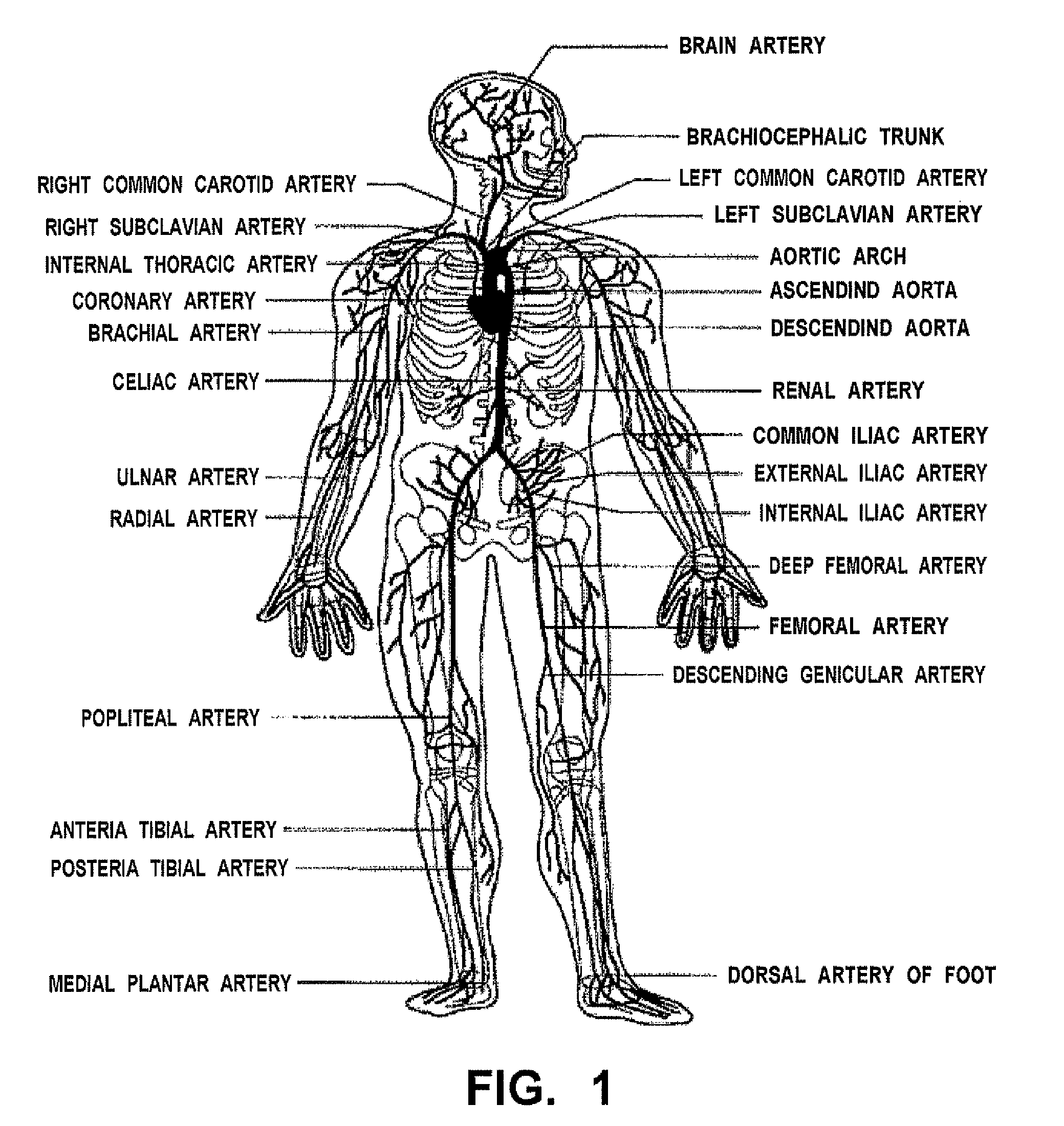
[0159]This artificial lesion member is arranged in a three-dimensional model (tube model) which is produced artificially by duplicating various kinds of tubes of a human living body provided, for example, with lumen portions such as a blood vessel (artery, vein), a lymphatic ...

second exemplified embodiment

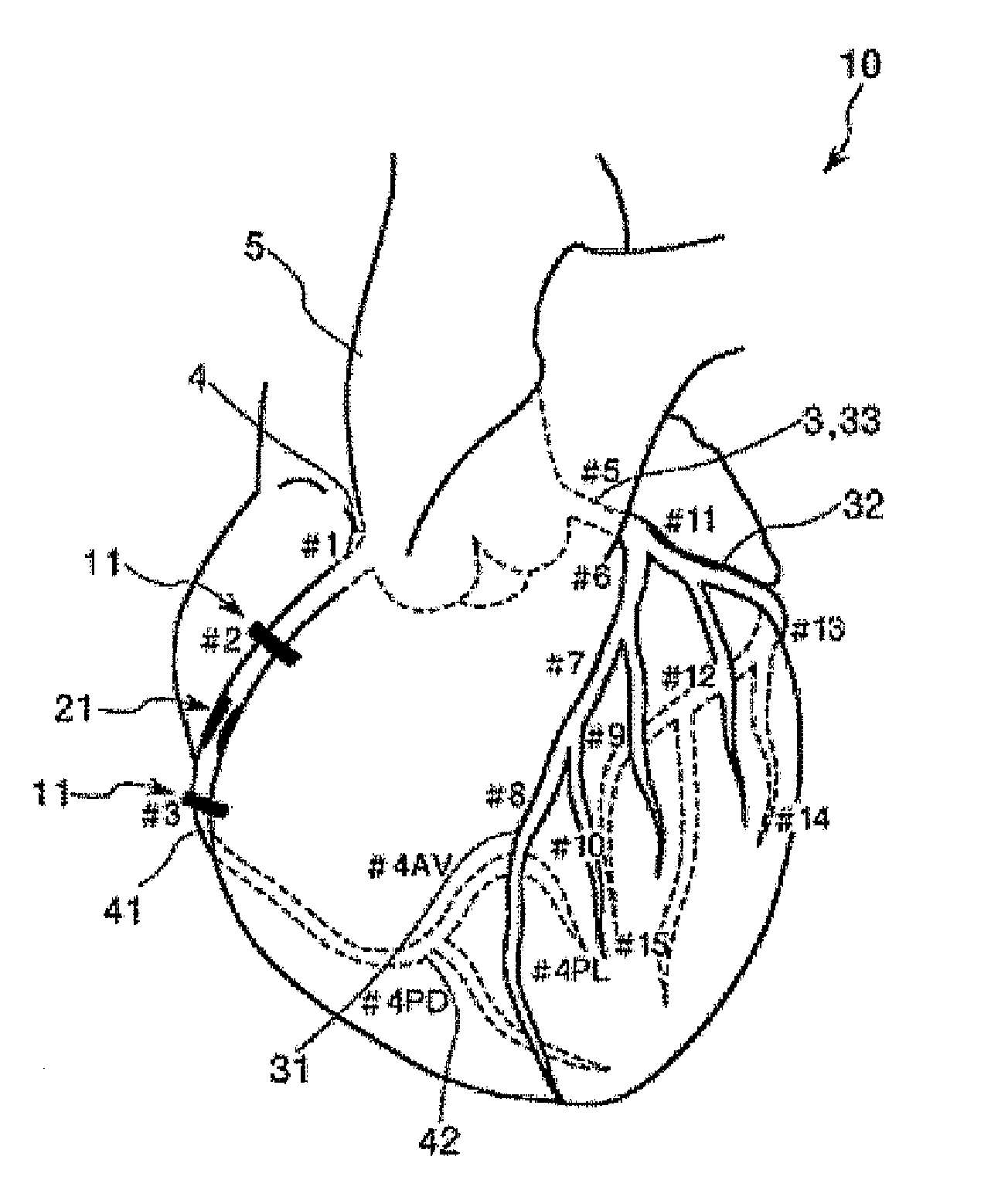
[0246]Next, it will be explained with respect to a second exemplified embodiment in which differently from the Segment 2 (#2: Middle) of the right coronary artery 4, there is arranged an artificial lesion member 21 on the left coronary artery 3.

[0247]Hereinafter, with respect to the second exemplified embodiment in which the artificial lesion member 21 is arranged in the left coronary artery 3, it will be explained centering around different aspects with respect to aforesaid first exemplified embodiment and with respect to similar matters, the explanation thereof will be omitted.

[0248]More specifically, in this exemplified embodiment (second exemplified embodiment), as shown in FIG. 15, it happens that there is employed a constitution similar to that of aforesaid first exemplified embodiment other than a fact that an artificial lesion member 21 is arranged at a branch portion (bifurcation) 34 in which Segment 6 of the left coronary artery 3 is branched into Segment 7 and Segment 9, ...

third exemplified embodiment

[0260]A biological model for training 1 of this exemplified embodiment includes the right coronary artery 4 (Segment 2) of the coronary artery 10, an artificial lesion member 121 arranged at the right coronary artery 4, and connection portions 11 provided respectively at both the end portions of the right coronary artery 4. For the right coronary artery 4, end portions of Segment 2 are respectively connected to Segment 1 and Segment 3 through the respective connection portions 11. In this case, it is preferable that the connection portions 11 are constituted so as to be freely detachably with respect to the Segment 1 and the Segment 3 respectively.

[0261]First, it will be explained with respect to the right coronary artery 4 which becomes Segment 2.

[0262]As shown in FIGS. 18A to 18B (similarly with respect to FIGS. 19A to 19D to FIGS. 22A to 22C), the right coronary artery 4 is an artery constituted by a tube shaped body 40 having a lumen portion 43. For the right coronary artery 4, ...


PUM


Abstract

Disclosed is a biological model for training which is in a tubular shape having an inner cavity, has a narrowing or closing portion at a mid portion of the tube, and is provided with a artificial lesion member simulating a lesion occurring in a tubular tissue. In this biological model for training, the artificial lesion member includes, at least in a portion thereof, a plastic and deformable material. The biological model for training is used in expansion training for implementing expansion. As the plastic and deformable material, a material including, as the main component, at least one member selected from among a silicone clay, a resin clay and an oil clay can be specifically used. Such a material is preferred because of ensuring the maintenance of a expanded state in the expansion training.

Description

TECHNICAL FIELD[0001]The present invention is an invention relating to a biological model for training and a production method of a biological model for training.BACKGROUND ART[0002]For one of percutaneous coronary angioplasties, there has been known, for example, a PTCA (Percutaneous Transluminal Coronary Angioplasty) operation.[0003]In this PTCA operation, in case of applying a method via femoral artery, blood flow inside a blood vessel is restored by way of the following procedures. More specifically, first, a sheath catheter is inserted into a femoral artery; subsequently, a guide wire for guide catheter is inserted thereinto; the guide catheter is advanced along the guide wire for guide catheter in a state of advancing the distal end thereof until the vicinity of a coronary artery entrance; and the distal end thereof is positioned at the coronary arterial orifice. <II> Next, the guide wire for guide catheter is pulled out; a guide wire for balloon catheter is inserted in...

Claims


Application Information

Patent Timeline
Patent Type & Authority Patents(United States)
IPC IPC(8): G09B23/28
CPCG09B23/285G09B23/30
Inventor MISAWA, HIROSHIKUCHIURA, TAKASHI
Owner TERUMO KK
Why Eureka
Try Eureka