
Magnetic resonance method and apparatus for triggered acquisition of magnetic resonance measurement data

a magnetic resonance and measurement data technology, applied in the field of magnetic resonance method and apparatus for triggered acquisition of magnetic resonance measurement data, can solve the problems of pathological changes, blurring, intensity loss and registration errors between images in reconstructed mr images, and being overlooked, so as to prolong the duration of the preparation module and increase the specific absorption ra

Active Publication Date: 2014-09-09
SIEMENS HEALTHCARE GMBH
View PDF6 Cites 2 Cited by
  • Summary
  • Abstract
  • Description
  • Claims
  • Application Information

AI Technical Summary

Benefits of technology

This approach significantly enhances the quality of unwanted signal suppression, ensuring consistent and effective reduction of unwanted signals in all acquired data, improving diagnostic accuracy by maintaining a steady state of unwanted tissue components before data acquisition.

Problems solved by technology

In magnetic resonance imaging (MRI) of examination regions affected by breathing movement, for example the organs of the thorax and abdomen, the breathing movement can lead to ghosting, blurring, intensity loss and registration errors between images in the reconstructed MR images.
These artifacts hinder the identification of findings by a physician on the basis of these MR images and can lead to pathological changes (such as lesions, for example) being overlooked.
However, the same or, respectively, a similar problem exists in the suppression of other unwanted signal components or in the suppression of fat with other methods.
Fat delivers a very intensive signal (for example in T2-weighted turbo spin echo images) that can outshine other signals and thus can hinder the finding [assessment] of various illnesses.
The shifted fat image interferes with the image impression and can superimpose lesions.
The fat suppression in the acquisition modules that are executed after the first preparation modules of the entire sequence is consequently not ideal, meaning that fat is not sufficiently suppressed.
In a magnetic resonance measurement that is not respiratory-triggered, this problem exists only once at the beginning of the measurement.
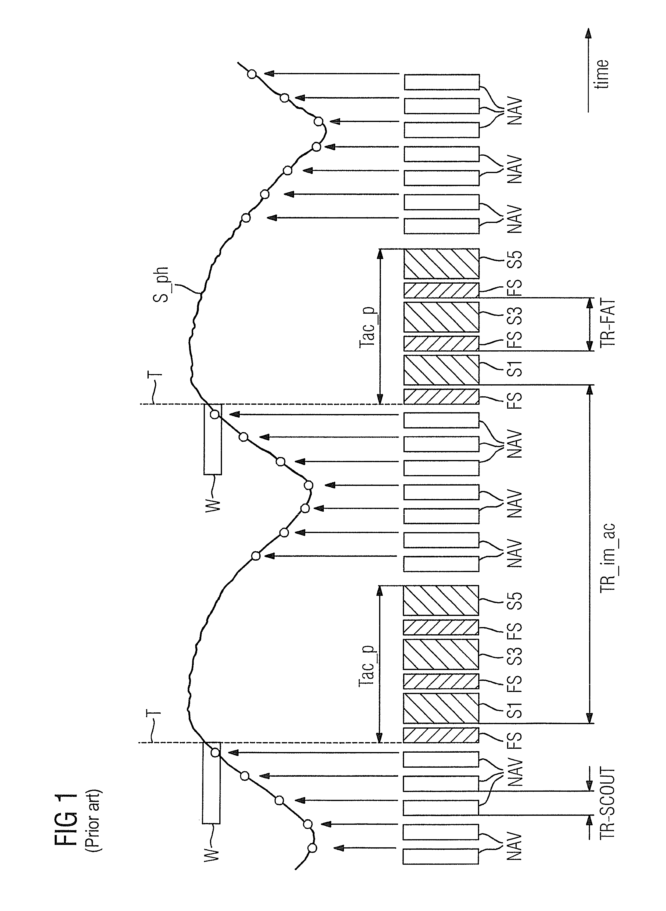
In particular, all data of a slice (and consequently the MR images that are calculated from these data) whose acquisition modules are executed relatively early after a trigger (slice S1 in FIG. 1, for example) are insufficiently fat-saturated.
This can significantly hinder the diagnosis with the aid of these images.
The problem illustrated above of the insufficient suppression of unwanted signals accordingly exists predominantly in techniques with respiratory triggering, but not in techniques which use respiratory gating, since there measurement can take place continuously (or quasi-continuously in the case of an EKG-controlled measurement).
However, in practice this does not work sufficiently well.

Method used


Image

  • Magnetic resonance method and apparatus for triggered acquisition of magnetic resonance measurement data
  • Magnetic resonance method and apparatus for triggered acquisition of magnetic resonance measurement data
  • Magnetic resonance method and apparatus for triggered acquisition of magnetic resonance measurement data

Examples


Embodiment Construction

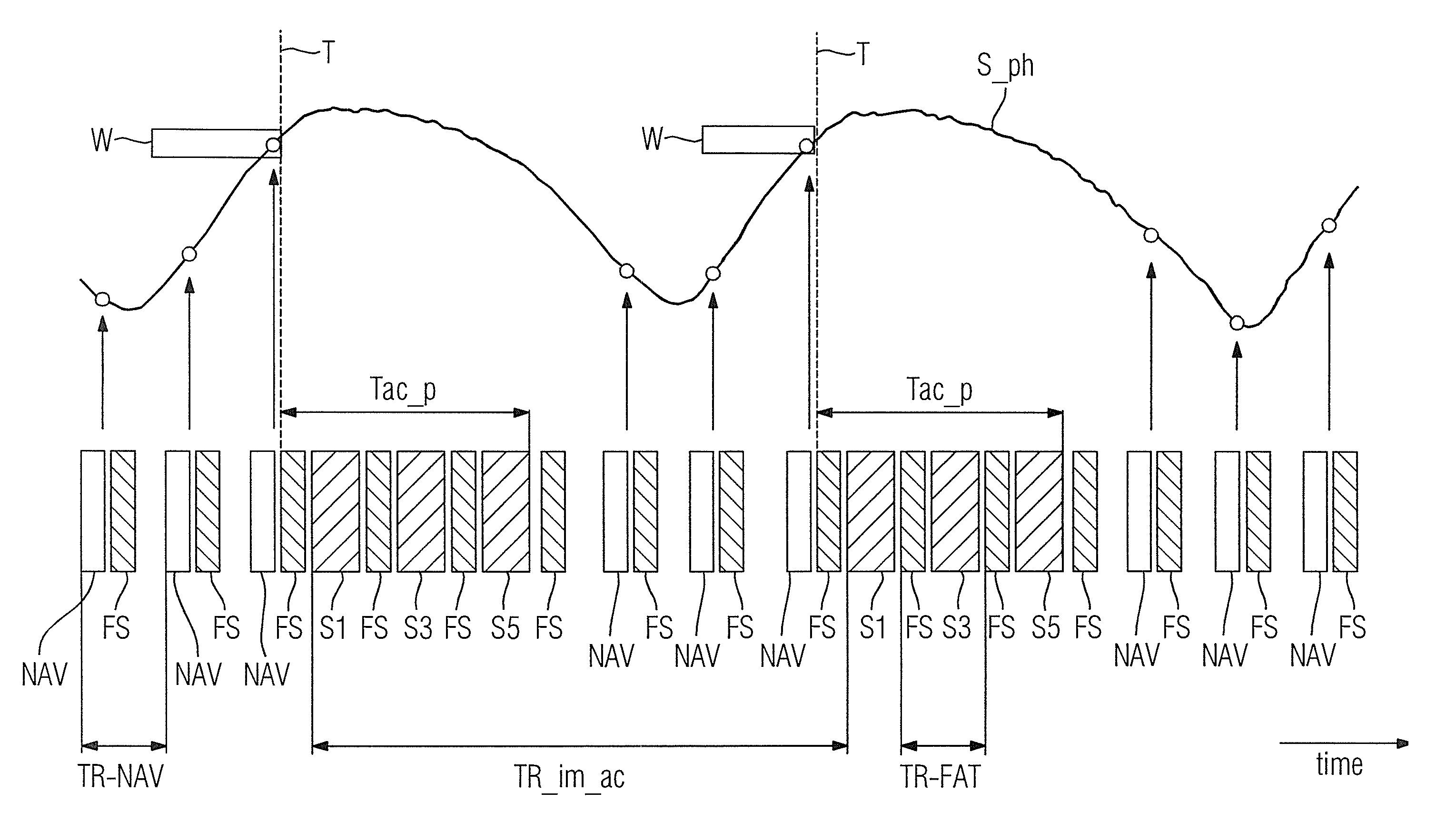
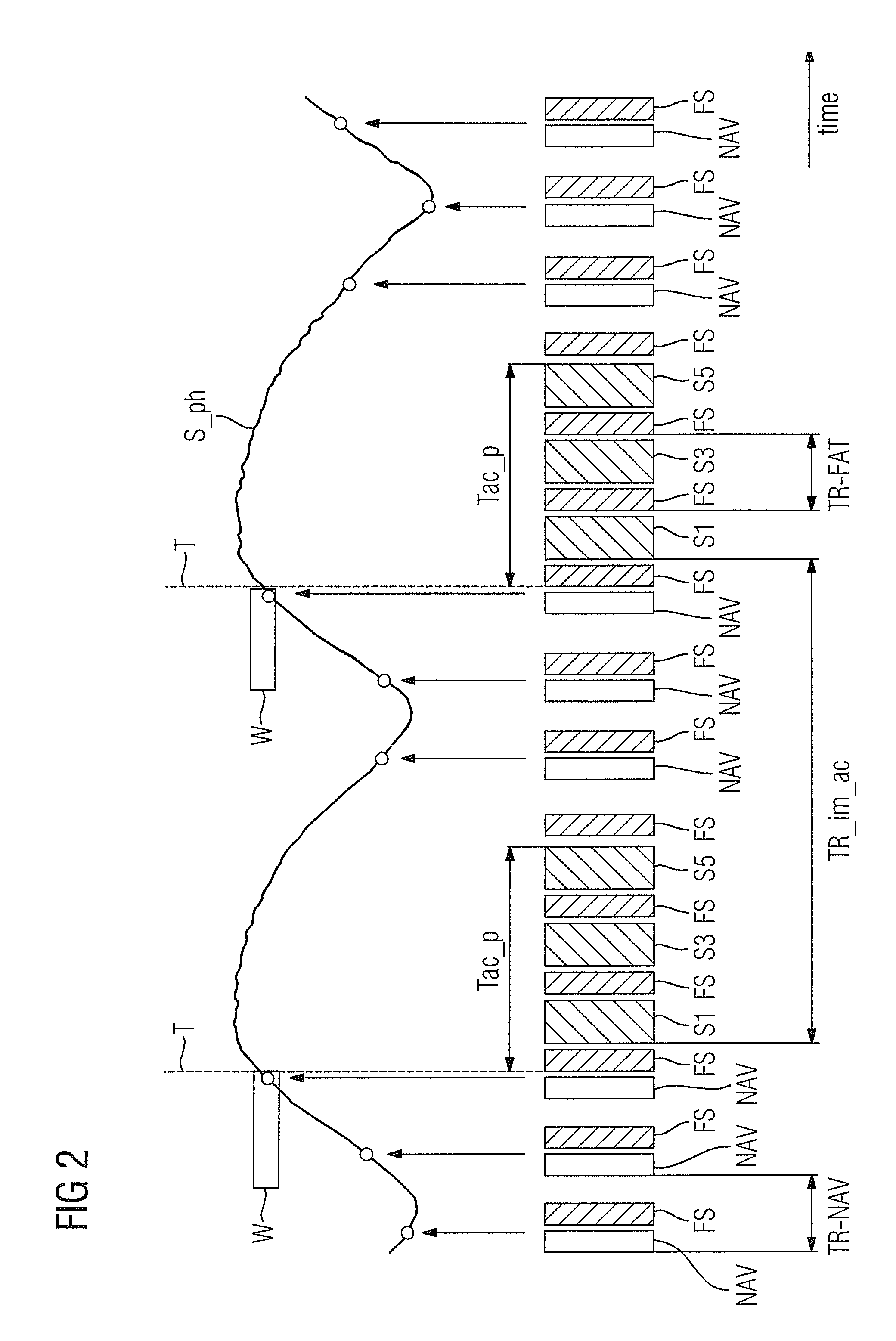
[0054]FIG. 1 schematically shows the chronological workflow of an example of a sequence of a respiratory-triggered MR measurement. A navigator sequence NAV is initially repeated with constant time interval TR-SCOUT. The result of each navigator measurement is a physiological data point (represented as a circle) that, for example, corresponds to a diaphragm position and thus reflects the breathing movement. The solid line S_ph that connects the measured data points (circles) serves to better visualize the underlying movement and therefore is shown solid, although only the measured data points shown as circles are actually present in the examination. Instead of the detection of the physiological data points by means of what is known as a navigator sequence, the physiological signal (here: breathing signal) can also be detected by means of an external sensor (for example a breathing cushion or a breathing belt) that can be connected with the magnetic resonance apparatus. In this case, ...


PUM


Abstract

In a magnetic resonance method and apparatus, a) data points of a physiological signal are detected, b) a trigger condition is evaluated depending on the detected physiological data points, c) a preparation module is executed to suppress unwanted signals in the time period in which the trigger condition has not yet been satisfied, d) after satisfying the trigger condition, an acquisition phase of predetermined duration is started, that includes at least two similar preparation modules to suppress unwanted signals and a respective following acquisition module to acquire measurement data, and e) after the acquisition phase, a) through d) are repeated until all desired measurement data have been acquired, with a time interval between two successive preparation modules being the same after a first execution of a preparation module in c) until the end of the acquisition phase in a subsequent d).

Description

BACKGROUND OF THE INVENTION[0001]1. Field of the Invention[0002]The present invention concerns a method to suppress unwanted signal components during an acquisition (triggered via a physiological signal of the examination subject) of magnetic resonance measurement data from an examination subject and a corresponding magnetic resonance apparatus, and a non-transitory computer-readable data storage medium to implement such a method.[0003]2. Description of the Prior Art[0004]The MR technique (MR: magnetic resonance) is a technique known for a few decades with which images of the inside of an examination subject can be generated. As a significantly simplified description, the examination subject is positioned in a comparably strong, static, homogeneous basic magnetic field (field strengths from 0.2 Tesla to 7 Tesla or more) in a magnetic resonance apparatus so that nuclear spins in the subject orient along the basic magnetic field. Radio-frequency excitation pulses are radiated into the...

Claims


Application Information

Patent Timeline
Patent Type & Authority Patents(United States)
IPC IPC(8): A61B5/055A61B5/00A61B5/08G01R33/56G01R33/567
CPCA61B5/7285G01R33/5607A61B5/055A61B5/0816G01R33/5676
Inventor STEMMER, ALTO
Owner SIEMENS HEALTHCARE GMBH
Learn More