Device for mobile electrocardiogram recording

a technology for electrocardiograms and devices, applied in the field of mobile medical diagnostic equipment, can solve the problems that the art does not teach or suggest such a device, system or method, and achieve the effects of reducing the need, improving the diagnostic ability of the device, and being well-rounded

Inactive Publication Date: 2014-12-02
CARDICELL
View PDF14 Cites 39 Cited by
  • Summary
  • Abstract
  • Description
  • Claims
  • Application Information

AI Technical Summary

Benefits of technology

[0019]The present invention overcomes these disadvantages of the background art by providing a device, system and method for obtaining a 12 lead electrocardiogram (ECG) from measurements obtained with 3 electrodes in some embodiments (it should be noted that some separate embodiments of the present invention relate to such measurements with 4 electrodes). Optionally and preferably, the device, system and method of the present invention may be easily and accurately operated by a layperson. Hereinafter, the term “layperson” refers to any individual without any substantial medical training.
[0034]Preferably, two electrodes are located on one face of the unit, so that one electrode is in contact with one arm and the other electrode is in contact with the other arm. A second face of the unit preferably includes one electrode (the third electrode) which is preferably initially in contact with the user's lower torso or leg. By placing the electrodes as described above, a recordation of the limb leads is preferably performed. In order to record the precordial limbs, the unit is then preferably moved to the user's upper torso. The hands are still preferably in contact with each of the two electrodes (located on the first face), but the third electrode (located on the second face) is now preferably in contact with the upper torso. By now moving the unit across the chest, each location V1 through to V6 is preferably in contact with the third electrode in turn (again, these locations are given as examples only without any intention of being limiting, as the device may optionally monitor electro-cardiac signals from all over the upper torso area, and is not necessarily limited only to the traditional V1 to V6 locations). Optionally, the potentials monitored from the limb leads may be stored in the device and are then preferably combined with the recorded V1 to V6 locations to further increase the accuracy of the precordial leads calculation.
[0070]The device of the present invention preferably comprises memory. The memory may optionally store medical information, and is preferably used to store medical history, most preferably including information regarding the cardiac history and status of the user (including but not limited to catheterization results, prior ECG's, echo results, stress test results and the like). The data can optionally be stored as video (for example for an angiogram, echocardiogram etc), images (for example CT, MRI), audio information and so on. Comparing the current ECG recording to the previously obtained ECG, which is stored in the device's memory, may increase specificity and sensitivity of diagnosis of cardiac abnormalities. Additionally the user's medical history may optionally and preferably be accessed in case of an emergency by medical personnel and may also be used for identifying the specific user. Optionally this memory also stores the upper torso locations as described above.
[0079]The device of the present invention may provide the option of performing many additional cardiac diagnostic tests some of which may require continuous use, for example including but not limited to performing a continuous type of monitoring known as a Holter test, or a stress test. Without wishing to be limited in any way, it should be noted that the test as described herein with the present invention may optionally be performed in a setting that is convenient and comfortable to the user, eliminating or at least reducing the need to perform these tests in a hospital setting. Such a test could optionally be performed as a “self” stress test which the user performs according to one or more instructions from the software.
[0080]Optionally the device may include communication with one or more other accessories (whether external or internal), such as a Pulse Oximeter, stethoscope, ultrasound transducer, echocardiogram transducer, thermometer, capnograph, blood pressure cuff, fetal heart rate transducer, camera, defibrillator electrodes, wire electrodes, inhaler, cardiac enzyme detector, blood sugar level analyzer and so forth. These accessories may, alternatively, be connected to the device optionally through one of the I / O plugs or wirelessly. The additional accessories may improve the diagnostic ability of the device and provide a well rounded depiction of the user's state of health.
[0081]The above mentioned ultrasound transducer may utilize the device's navigation system in order to reach the conventional ultrasound locations. The ultrasound may monitor different heart functions (such as the motion of the walls, the valve functions etc.). The device is preferably able to compare between the stored ultrasound parameters and the acquired real time parameters which for increasing the sensitivity of detection of heart abnormalities. A stethoscope may optionally be used in a similar manner for locating the monitoring points using the navigation system.

Problems solved by technology

The background art also does not teach or suggest such a device, system or method which may be easily and accurately operated by a layperson (without any substantial medical training).
The background art also does not teach or suggest such a device, system or method which is mobile and which is able to provide results to a remote location, for example to medical personnel located at such a remote location from the subject undergoing the ECG measurements.

Method used


Image

  • Device for mobile electrocardiogram recording
  • Device for mobile electrocardiogram recording
  • Device for mobile electrocardiogram recording

Examples


Embodiment Construction

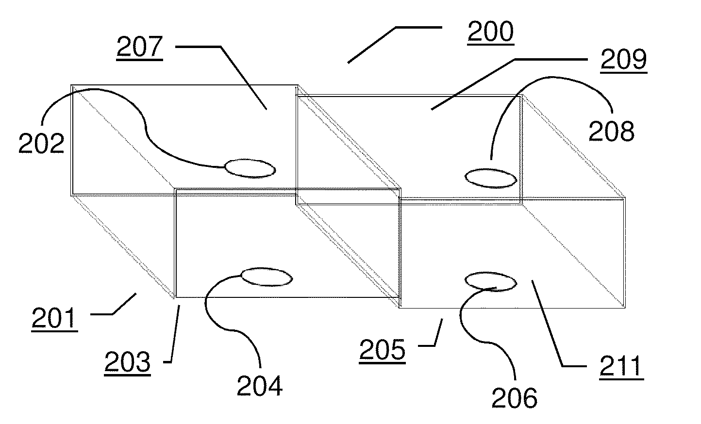
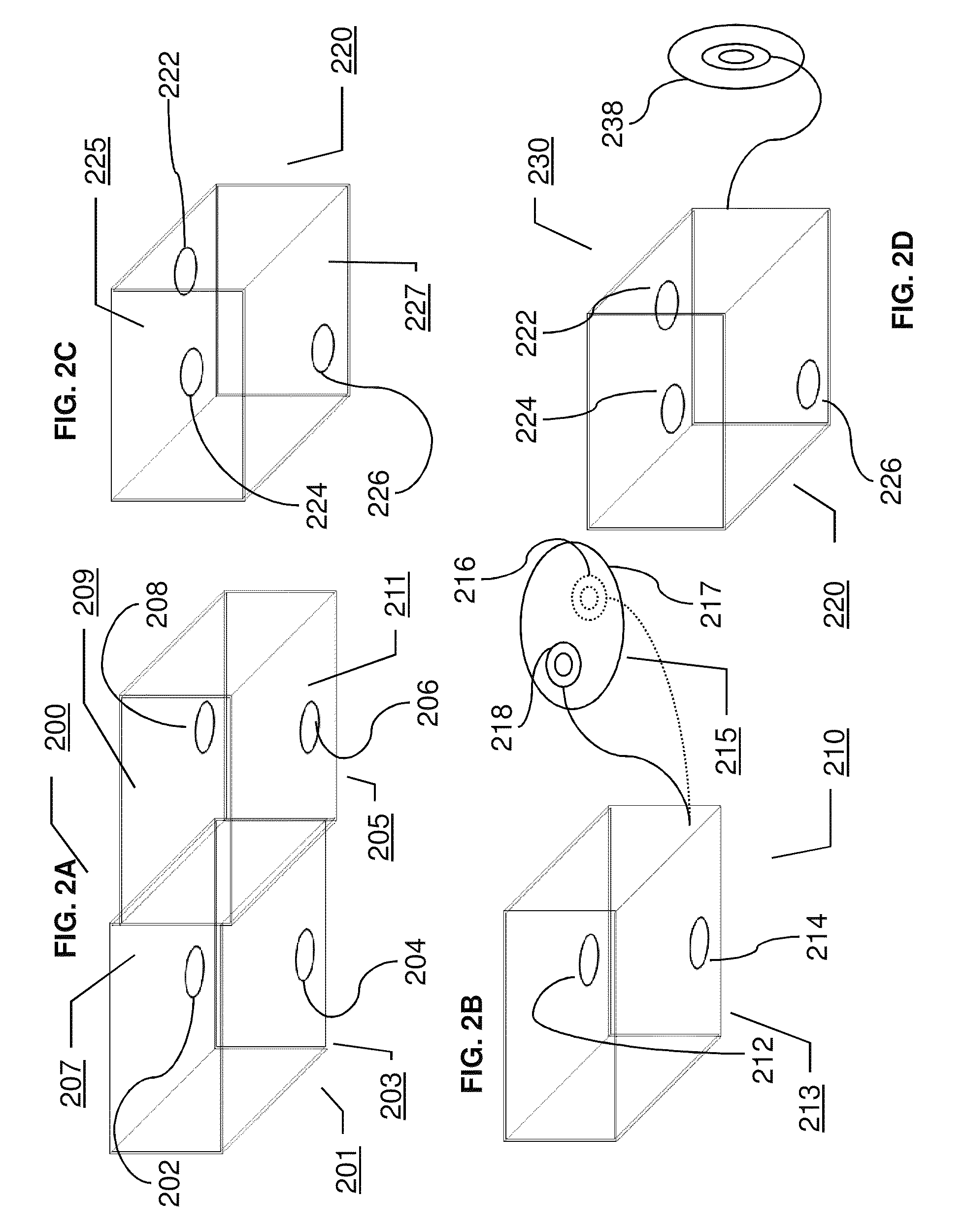
[0174]The present invention is of a system, personal device and a method for recording a 12 lead ECG and / or other biological signals by using a small number of electrodes. According to some embodiments, three electrodes are used while according to other embodiments, four electrodes are used. Each electrode is preferably in contact with a different body part. Optionally, the electrodes are contained in one unit but preferably the electrodes are divided between two units. More preferably, the two units communicate with each other, optionally through wires for example.

[0175]The electrodes may optionally be distributed between the units (if more than one is present) according to any suitable configuration, including the exemplary configurations described herein. Regardless of the exact configuration, according to some embodiments, a method for obtaining a 12 lead ECG reading from three or four electrodes preferably includes obtaining at least one measurement from a location selected fro...


PUM


Abstract

A device, system and method for obtaining a 12 lead electrocardiogram (ECG) from measurements obtained with 3 electrodes in some embodiments (it should be noted that some separate embodiments of the present invention relate to such measurements with 4 electrodes). Optionally and preferably, the device, system and method of the present invention may be easily and accurately operated by a layperson.

Description

[0001]This Application is a national phase of, and claims priority from, PCT Application No. PCT / IL2007 / 000947, filed on Jul. 29 2007, which claims priority from U.S. Provisional Application No. 60 / 820,780, filed on Jul. 29 2006, all of which are hereby incorporated by reference as if fully set forth herein.FIELD OF THE INVENTION[0002]The present invention relates to a device, system and a method for a mobile medical diagnostic equipment and in particular, to such a system and method for an electrocardiogram.BACKGROUND OF THE INVENTION[0003]Heart problems are one of the leading causes of morbidity and mortality worldwide.[0004]Ischemic heart disease and arrhythmias are the two major types of cardiac abnormalities with prevalence of 12% in the general population, manifested in a wide range of symptoms, most typically chest pain radiating to other parts of the body.[0005]Chest pain is a common symptom occurring in many diseases other than heart disease and since acute ischemic or arrh...

Claims


Application Information

Patent Timeline
Patent Type & Authority Patents(United States)
IPC IPC(8): A61B5/04A61B5/0408A61B5/0428A61B5/06A61B5/00A61B5/0402A61B5/308
CPCA61B5/04286A61B5/04085A61B5/061A61B5/6831A61B5/04028A61B5/684A61B5/327A61B5/282A61B5/303
Inventor BERKNER, LIOR
Owner CARDICELL
Why Eureka
Try Eureka