Intramedullary fixation device for small bone fractures

a technology for fixing devices and fractures, applied in the field of small bone fracture fixation devices, can solve the problems of not having the memory curve, not being able to perform surgery, and not being able to straighten k-wires again, and achieve the effects of increasing the arc of curvature, and increasing the bending or flexion

Inactive Publication Date: 2014-12-23
MAYO FOUND FOR MEDICAL EDUCATION & RES
View PDF42 Cites 2 Cited by
  • Summary
  • Abstract
  • Description
  • Claims
  • Application Information

AI Technical Summary

Benefits of technology

[0011]The present invention addresses the foregoing needs by providing a system for the fixation of fractures of small bones such as phalangeal, metacarpal, and metatarsal bones. The system uses curved shape-memory alloy (e.g., Nitinol) wires that have a predetermined radius of curvature to accommodate different sized bones. Multiple sizes of the wires can be included in a kit. These shape-memory alloy wire forms are inserted into phalanx, metacarpal or metatarsal bones via a percutaneous technique. The technique uses small skin incisions; a specialized drill guide that has holding K-wires to maintain fixation of the drill guide to the bone so that it does not lose the insertion point; a specialized drill as well as a specialized wire cutter and advancement tool to make sure that the level of the wire is below the level of the outer cortical bone. Shape-memory alloy (e.g., Nitinol) based wires with a pre-bent curve have an advantage over the typical standard K-wire in that they can be allowed to spring back to their predetermined shape when inserted into the intramedullary canal of the bone and heated, i.e., a more aggressive curve. By increasing their bending or flexion to increase the arc of curvature, this allows fixation points for the wire form, essentially locking it to bone. This solves a problem with typical straight K-wire fixation that does not have locking points that prevent movement or migration of the wire-bone construct.
[0013]Also, the use of a specialized drill prevents over penetrating of the bone. This drill also has a specialized tip that allows it to catch on the bone preventing the usual movement of the drill once it starts because of the torque from the drill bit forcing the drill to move off of the bone. A shoulder ledge on the drill is used to prevent over penetration. Once the bone has essentially been close reduced and the drill guide placed through a small incision in the drill used to penetrate the cortex, the intramedullary device is advanced in through a hand toggling system. This can be checked under fluoro-imaging to make sure that it was advancing appropriately across the fracture site. It is advanced only up to a certain point so that the wire can be then further advanced once it had been cut to fit completely in the bone. Once the wire is imaged and perceived to be in the appropriate position, the shape-memory alloy is heated to restore its memory and increase its curvature points. The guide is then reduced and a special wire cutter cuts the intramedullary nail very close to the bone. The guide can then be reapplied along the same previous K-wires that were there and then a wire advancement tool used through the drill guide to push the remaining wire into the bone so that it sits at or below the level of the cortical surface. The wire advancement tool can have a special groove in the base of it to catch the bump or ridge left by the wire cutter to help facilitate maintaining some rotational control. An alternative means is to precut or cut prior to heating the metal. This then advances the intramedullary device into the bone again prior to the memory being activated. The memory can then be activated by drilling two specialized wire probes to touch two different sections of the intramedullary device and then applying the heating electrical charge through these two percutaneous wires that were then touching the shape-memory alloy in the bone.
[0014]Current methods for treating metacarpal and metatarsal fractures intramedullary with K-wires involve making an incision over the proximal aspect of the bone and drilling a hole in it large enough so that the wire can be advanced down the intramedullary canal. With shape-memory alloy intramedullary devices it is possible to insert this through the dorsal superior aspect of the metacarpal and metatarsal heads. This precludes the need for drilling a large hole into the bone. Instead this wire can be advanced and once it was passed the fracture site and engages the dorsal cortex, the memory can be activated, the wire cut at the level of the chondral bone and then again with the advancement tool, advanced another millimeter or two. It can be quite easy to insert, certainly one if not two of these wires down the intramedullary canal in this fashion providing a fixation not only for diaphyseal type metacarpal and metatarsal fractures, but for also the more challenging metacarpal neck fractures, i.e., boxer fracture. Boxer fractures in this country are quite common. However, they can be challenging to treat in that there usually is not adequate bone distally for a traditional plate and screw fixation and that K-wire fixation with proud wires sticking through the skin can potentially lead to deep infections of the joint or soft tissues, or a significant irritation with scarring and / or stiffness of the small finger itself. This invention can preclude these types of problems by providing intramedullary fixation, which allows for early active range of motion, improving patient outcomes.

Problems solved by technology

Shape-memory alloy (e.g., Nitinol) wires in suitable diameters are commercially available; however, they do not have the memory curve “programmed” into them and they are not surgical grade.
Straight K-wires again, have the disadvantage of acting only in an axial fashion.
However, they can be challenging to treat in that there usually is not adequate bone distally for a traditional plate and screw fixation and that K-wire fixation with proud wires sticking through the skin can potentially lead to deep infections of the joint or soft tissues, or a significant irritation with scarring and / or stiffness of the small finger itself.

Method used


Image

  • Intramedullary fixation device for small bone fractures
  • Intramedullary fixation device for small bone fractures
  • Intramedullary fixation device for small bone fractures

Examples


Embodiment Construction

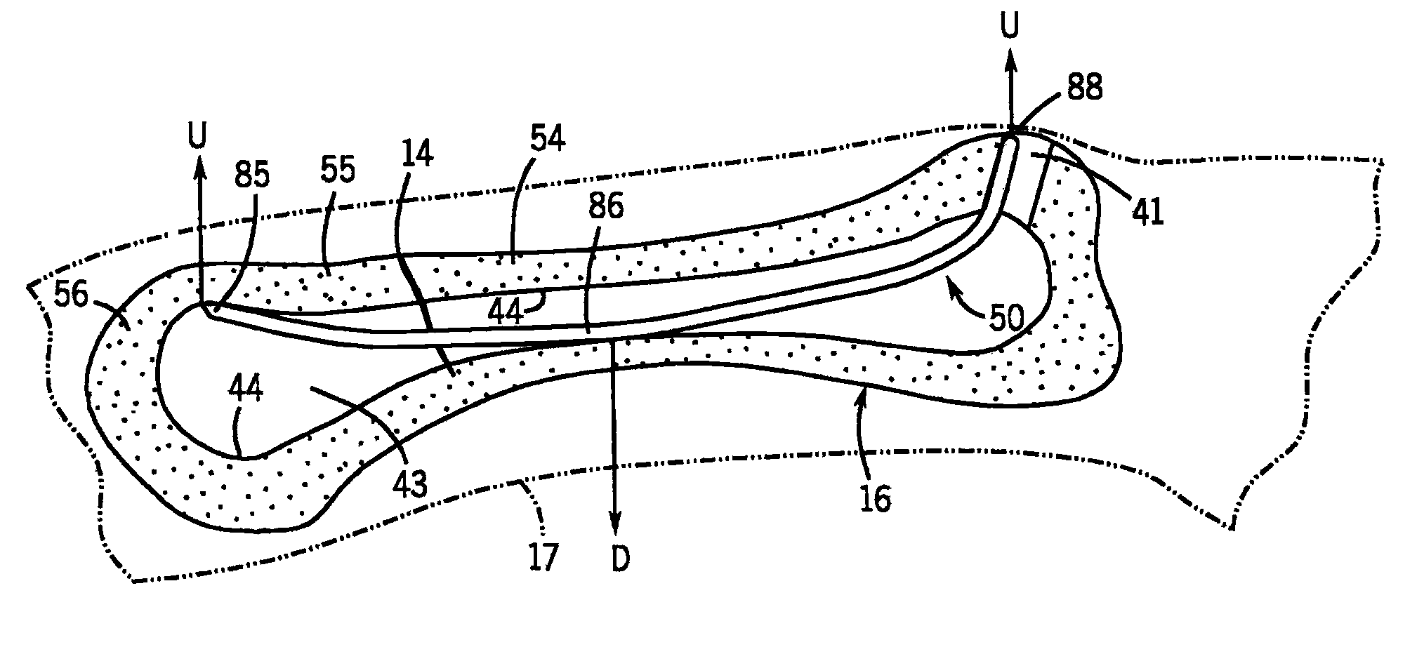
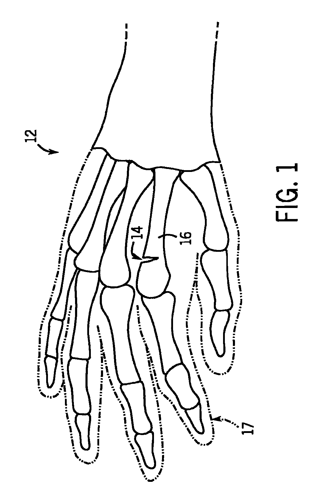
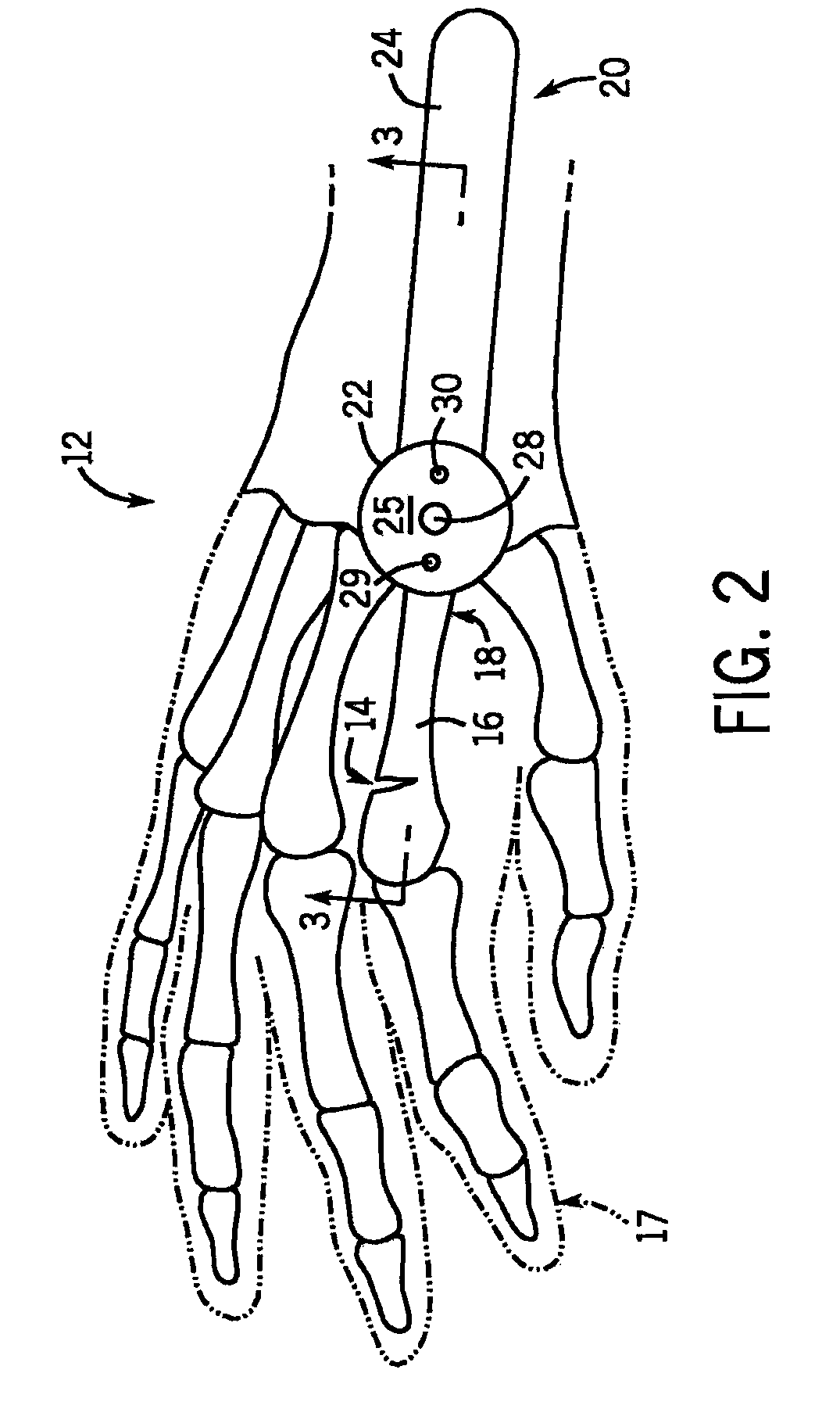
[0043]Looking first at FIG. 1, there is shown a right human hand 12 having a fracture 14 in the second metacarpal 16 in the index finger 17. The method and devices according to the invention can be used for fixation of the fracture 14 of the second metacarpal 16 to maintain proper fracture reduction for healing. While the use of methods and devices according to the invention has been shown and described herein with reference to a fracture 14 of the second metacarpal 16, it should be appreciated that the methods and devices according to the invention can be used for the fixation of other small bone fractures, such as fractures of any of the phalangeal, metacarpal, and metatarsal bones. Also, while the methods and devices according to the invention shown and described herein only use a single wire form, one or more wire forms may be inserted into the intramedullary canal of the bone in the method.

[0044]In the method of the invention, an incision is first made in the skin and tissue ov...


PUM


Abstract

A fracture fixation system particularly useful for bones of the hand and foot is disclosed. The system uses curved shape-memory alloy (e.g., Nitinol) wires that have a predetermined radius of curvature to accommodate different sized bones. These shape-memory alloy wire forms can be inserted into phalanx, metacarpal or metatarsal bones via a percutaneous technique. The technique uses small skin incisions; a specialized drill guide that has holding K-wires to maintain fixation of the drill guide to the bone so that it does not lose the insertion point; a specialized drill as well as a specialized wire cutter and advancement tool to make sure that the level of the wire is below the level of the outer cortical bone. Shape-memory alloy (e.g., Nitinol) based wires with a pre-bent curve have an advantage over the typical standard K-wire in that they can spring back to their predetermined memory shape when inserted into the intramedullary canal of the bone and heated, i.e., a more aggressive curve. By increasing their bending or flexion to increase the arc of curvature, this allows fixation points for the wire, essentially locking it to bone.

Description

CROSS-REFERENCES TO RELATED APPLICATIONS[0001]This application claims priority from U.S. Provisional Patent Application No. 61 / 050,386 filed May 5, 2008.STATEMENT REGARDING FEDERALLY SPONSORED RESEARCH[0002]Not Applicable.BACKGROUND OF THE INVENTION[0003]1. Field of the Invention[0004]The invention relates to methods and devices for the fixation of small bone fractures, and more particularly to methods and devices for the fixation of fractures of the phalangeal, metacarpal, and metatarsal bones.[0005]2. Description of the Related Art[0006]Bones which have been fractured should be immobilized on either side of the fracture for proper healing. Fractures of the phalanges and metacarpals of the hand, and the phalanges and metatarsals of the foot are quite common. Various techniques and fixation devices have been used to maintain proper fracture reduction in the phalangeal, metacarpal, and metatarsal bones.[0007]One fixation method for phalangeal, metacarpal, and metatarsal bones uses pl...

Claims


Application Information

Patent Timeline
Patent Type & Authority Patents(United States)
IPC IPC(8): A61B17/68A61B17/92A61B17/17A61B17/72A61B17/00
CPCA61B2017/1782A61B2017/1775A61B17/921A61B17/1725A61B2017/00867A61B17/72A61B17/1775A61B17/1782
Inventor DUNCAN, SCOTT, F., M.
Owner MAYO FOUND FOR MEDICAL EDUCATION & RES
Why Eureka
Try Eureka