Process, system and software arrangement for measuring a mechanical strain and elastic properties of a sample

a technology of elastic properties and mechanical strain, applied in the field of measuring mechanical strain and elastic properties of samples, can solve the problems of high-risk scenario, insufficient knowledge of structural and compositional features alone, and inability to accurately predict plaque rupture, etc., to achieve the reduction of parameter search space, the effect of preserving sharp spatial gradients in strain or modulus, and minimizing partial volum of tissue types within each elemen

Active Publication Date: 2015-02-24
THE GENERAL HOSPITAL CORP
View PDF641 Cites 20 Cited by
  • Summary
  • Abstract
  • Description
  • Claims
  • Application Information

AI Technical Summary

Benefits of technology

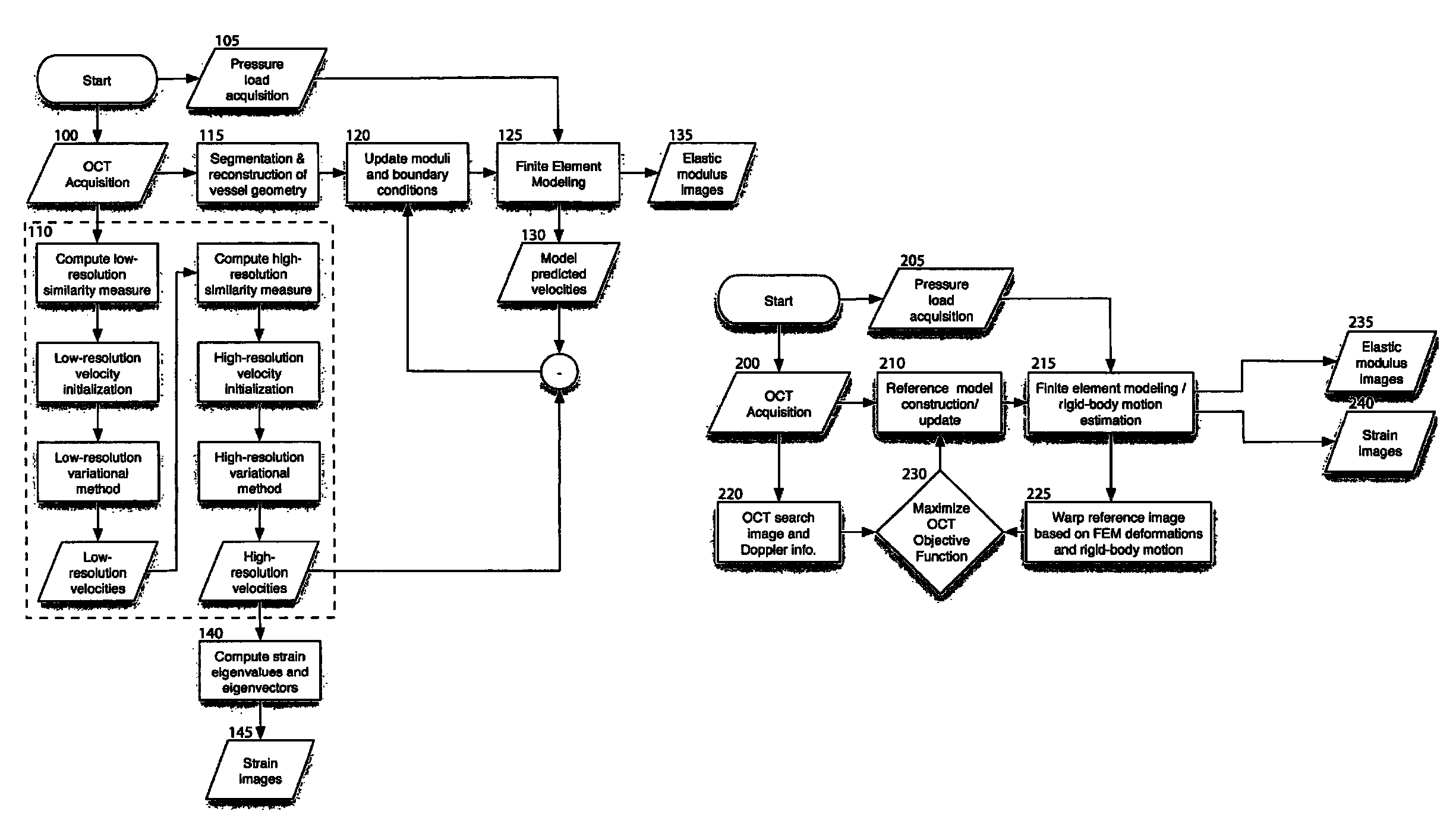
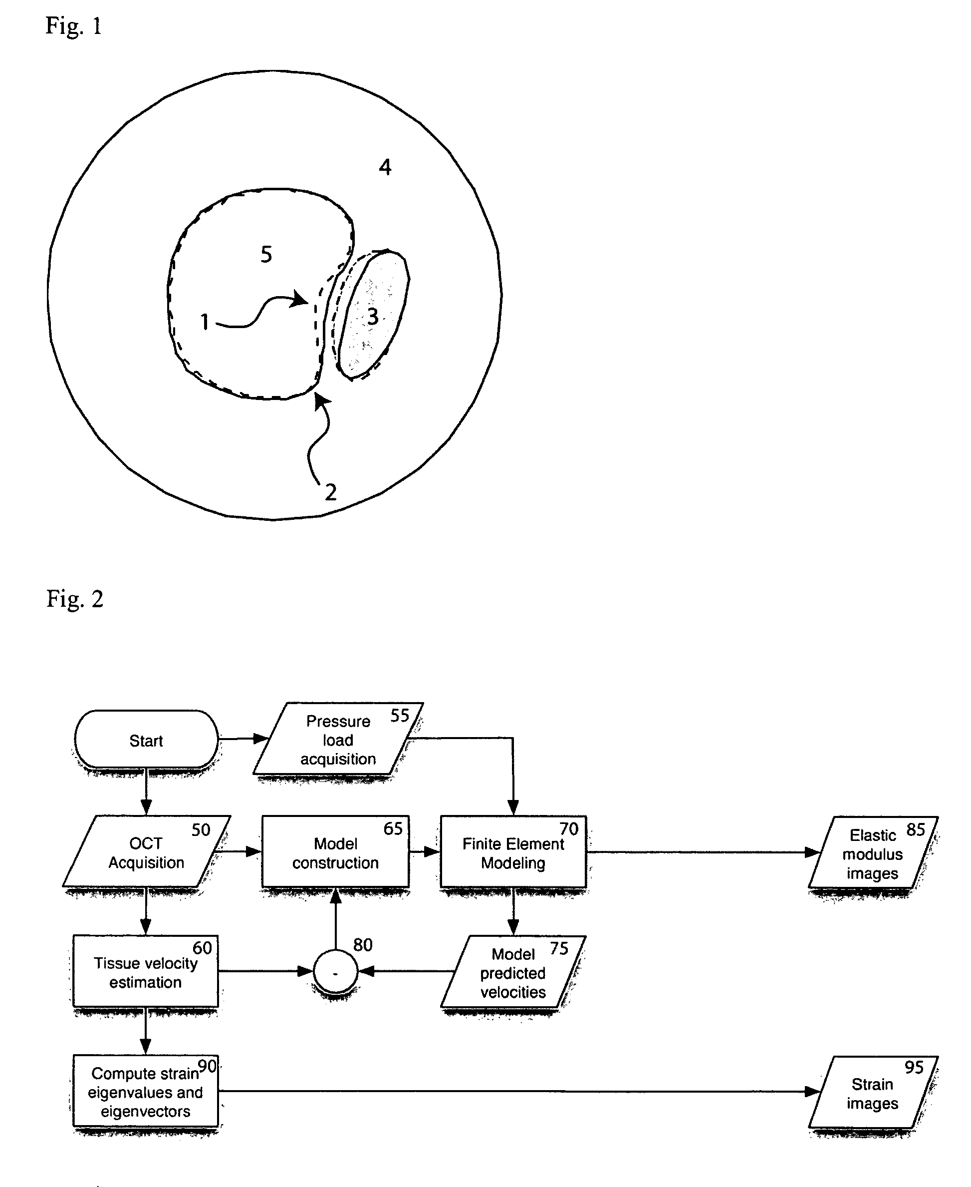
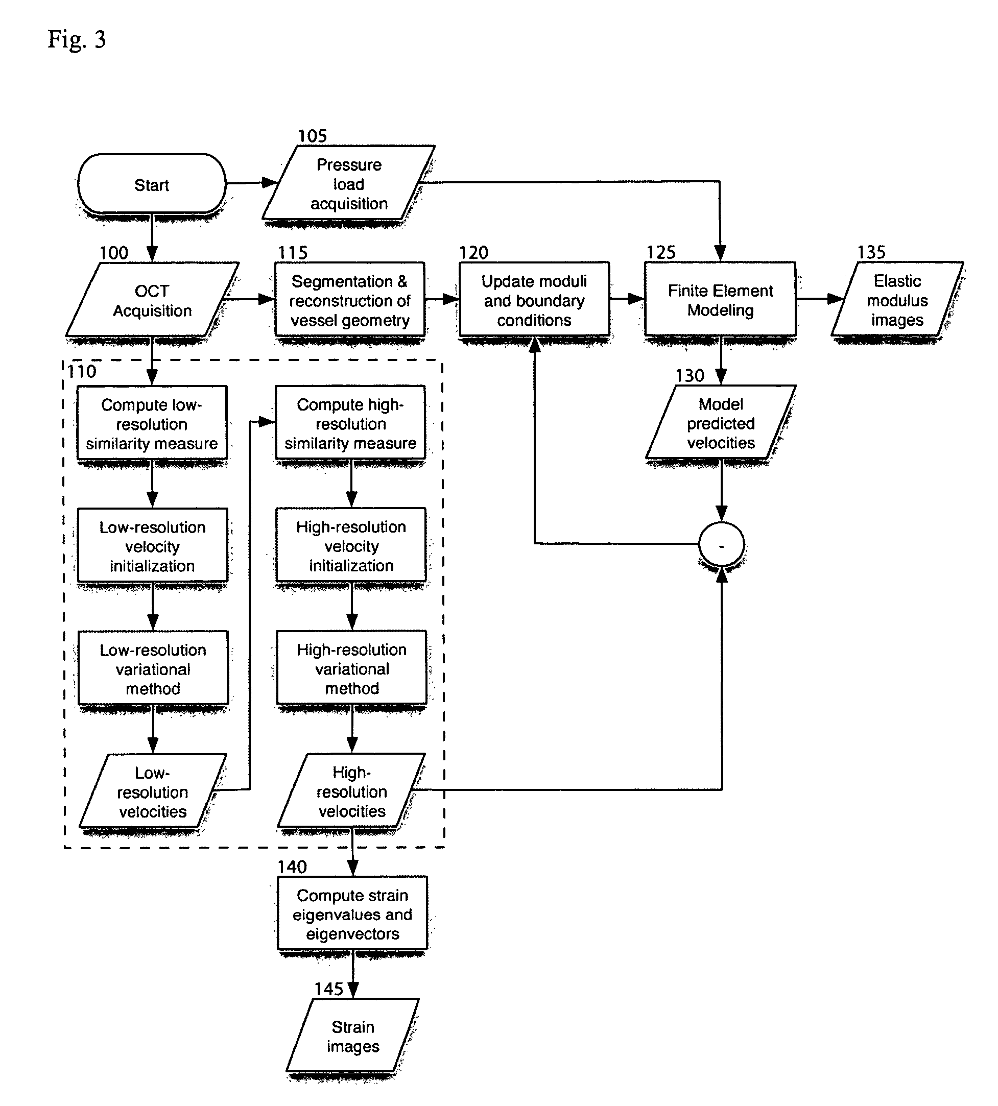
[0015]In one exemplary embodiment of the present invention, OCT can be used to determine the structure and tissue composition of a vessel. This information may then be used to construct a numerical model representing the vessel and finite element modeling, using estimates of elastic moduli, can be subsequently used to predict the mechanical response of the vessel to a given stress load. Separately from this exemplary computation, an exemplary OCT elastography technique according to the present invention may be performed to measure the mechanical response of the vessel. The two pathways, modeling and imaging, can represent a) a prediction based on assumed elastic modulus distribution; and b) a measurement, respectively. The difference between these two results can be considered as an error function to be minimized by a modification of the initial estimate for the elastic modulus distribution. Through an iteration of this exemplary technique according to the present invention, the distribution and magnitude of elastic modulus can be determined. Such information could be displayed as a cross-sectional or three-dimensional image of elastic modulus. Additionally, by minimizing the error function, an improved elastographic image of strain can be generated. As a result, the exemplary embodiments of the system, process and software arrangement according to the present invention are capable of overcoming the limitations of current diagnostic technology wherein structure and / or strain are measured, and the biomechanical characteristics of the tissue remain unknown. Further, the present invention improves upon the resolution and sensitivity of previous methods for elastography.
[0023]Further, in contrast to conventional methods and techniques for biomechanical imaging, another exemplary embodiment of the present invention takes into consideration that tissues of the same type would likely have similar, and possibly almost identical, mechanical properties, and that high-tissue-contrast may be available in the OCT techniques for a segmentation of regions-of-interest into distinct tissue components (e.g., fibrous, lipid, calcified, etc.). This exemplary embodiment of the process according to the present invention can preserve the boundaries present between tissues, while reducing the parameter search space. For example, unlike estimation on a low-resolution grid, partial voluming of tissue types within each element can be minimized with this technique, allowing for sharp spatial gradients in strain or modulus to be preserved. In addition, an adaptive mesh refinement at elements where the biomechanical model poorly fits the data can be a contribution that is beneficial to the elastography and modulus imaging techniques.

Problems solved by technology

Myocardial infarction is a major cause of death in industrialized countries.
The combination of biochemically initiated weakening, represented by these three features and elevated mechanical stress, may represent a particularly high-risk scenario.
However, the knowledge of structural and compositional features alone may be insufficient for a detailed understanding and accurate prediction of plaque rupture.
Although these numerical techniques have provided some insight into the plaque rupture, they are disadvantageous because, e.g., a) their accuracy is limited by the imprecise knowledge of the elastic properties and their distribution; and b) they are based on retrospective data, and may not be directly applied to the assessment of the vascular structure in living patients.
Although this technique can be performed in vivo, it provides a low spatial resolution and low contrast between typical tissue components in the atherosclerotic plaques.
Further, such technique does not provide the ability to determine the stress independently from the strain, and therefore may not be capable of determining the elastic modulus distributions.
However, the technique for simultaneously determining structure, composition and biomechanical properties of a sample is not available.
As the parameter space grows, the search for parameter estimates that satisfy the desired objective functional becomes a difficult underdetermined problem.
In addition, large computational costs and computing time are generally incurred to probe parameter spaces of high-dimensionality (on the order of >100 dimensions).
These conventional methods can be time-consuming, requiring several minutes of processing for large regions of interest.

Method used


Image

  • Process, system and software arrangement for measuring a mechanical strain and elastic properties of a sample
  • Process, system and software arrangement for measuring a mechanical strain and elastic properties of a sample
  • Process, system and software arrangement for measuring a mechanical strain and elastic properties of a sample

Examples


example

[0113]The following description provides details on the experimental testing of an exemplary embodiment of the method according to the present invention: In particular, the exemplary multi-resolution variational method was performed in simulated OCT imaging during axial compression of a tissue block containing a circular inclusion. FIG. 6 depicts a finite element model geometry 300 and corresponding finite element mesh 305 used for the tissue block and circular inclusion. Sequential interference images were generated as described in equations (1)-(6) by computing the product of an exponential decay term and a convolution between the coherent OCT point-spread function and the distribution of backscattering arising from point scatterers moving in the sample.

[0114]Backscattering values at discrete points within the tissue block were simulated as independent uniform random variables with a variance of 10 for scatterers within the block and a variance of 2 for scatterers within the circu...


PUM


Abstract

A system, process and software arrangement are provided to determining data associated with at least one structural change of tissue. In particular, a first optical coherence tomography (“OCT”) signal which contains first information regarding the tissue at a first stress level, and a second OCT signal which contains second information regarding the tissue at a second stress level are received. The first and second information are compared to produce comparison information. The data associated with the at least one structural change is determined as a function of the comparison information and further information associated with (i) at least one known characteristics of the tissue and / or (ii) characteristics of an OCT system. Further, at least one optical coherence tomography (“OCT”) signal which contains information regarding the tissue can be received, and the modulus of the tissue may be determined as a function of the received at least one OCT signal.

Description

CROSS-REFERENCE TO RELATED APPLICATION(S)[0001]The present invention claims priority from U.S. Patent Application Ser. No. 60 / 604,137 filed on Aug. 24, 2004, the entire disclosure of which incorporated herein by reference.STATEMENT REGARDING FEDERALLY SPONSORED RESEARCH [0002]This invention was made with government support under Grant No(s). DAMD17-99-2-9001 awarded by the U.S. Army Medical Research and Material Command. The government has certain rights in the invention.FIELD OF THE INVENTION[0003]The present invention relates generally measuring a mechanical strain and elastic properties of a sample, and more particularly, to a process, system and software arrangement for non-invasively measuring and determining a spatial distribution of a mechanical strain and elastic properties of biological samples.BACKGROUND OF THE INVENTION[0004]Myocardial infarction is a major cause of death in industrialized countries. Rupture of vulnerable atherosclerotic plaques has been recognized as an ...

Claims


Application Information

Patent Timeline
Patent Type & Authority Patents(United States)
IPC IPC(8): A61B5/02A61B5/00
CPCA61B5/0066A61B5/0059A61B5/02007
Inventor BOUMA, BRETT EUGENECHAN, RAYMOND C.TEARNEY, GUILLERMO J.
Owner THE GENERAL HOSPITAL CORP
Why Eureka
Try Eureka