Determining electrical properties of tissue using magnetic resonance imaging and least squared estimate

a technology of magnetic resonance imaging and tissue electrical properties, applied in the direction of instruments, diagnostic recording/measuring, measurement using nmr, etc., can solve the problems of reducing the resolution of the image corresponding to the electrical properties, poor implementation of conventional approaches to generating electrical properties maps, and affecting the accuracy of the results

Active Publication Date: 2015-06-30
GENERAL ELECTRIC CO
View PDF9 Cites 7 Cited by
  • Summary
  • Abstract
  • Description
  • Claims
  • Application Information

AI Technical Summary

Problems solved by technology

Determining the electrical properties of tissue in-vivo using MRI has posed several problems due to the inability to directly measure the phase of the receive RF magnetic field B1− and the phase of the transmit RF magnetic field B1+.
While conventional approaches have provided techniques for estimating the electrical properties of tissue based on mapping the amplitude of B1+ and approximating the phase of B1+, implementations of conventional approaches to generating electrical properties maps are vulnerable to poor results due to noise in B1+ data.
Typically, the use of such skip factors requires a larger area for the estimation of the Laplacian at each pixel location, which can reduce the resolution of the images corresponding to the electrical properties.
These approaches can lead to lower resolution or inaccurate results.

Method used


Image

  • Determining electrical properties of tissue using magnetic resonance imaging and least squared estimate
  • Determining electrical properties of tissue using magnetic resonance imaging and least squared estimate
  • Determining electrical properties of tissue using magnetic resonance imaging and least squared estimate

Examples


Embodiment Construction

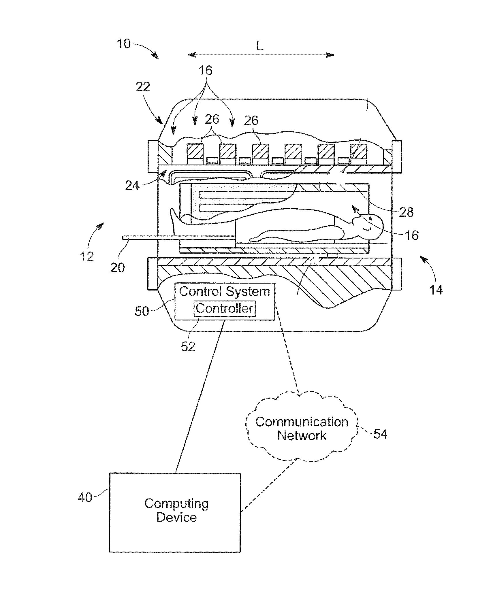
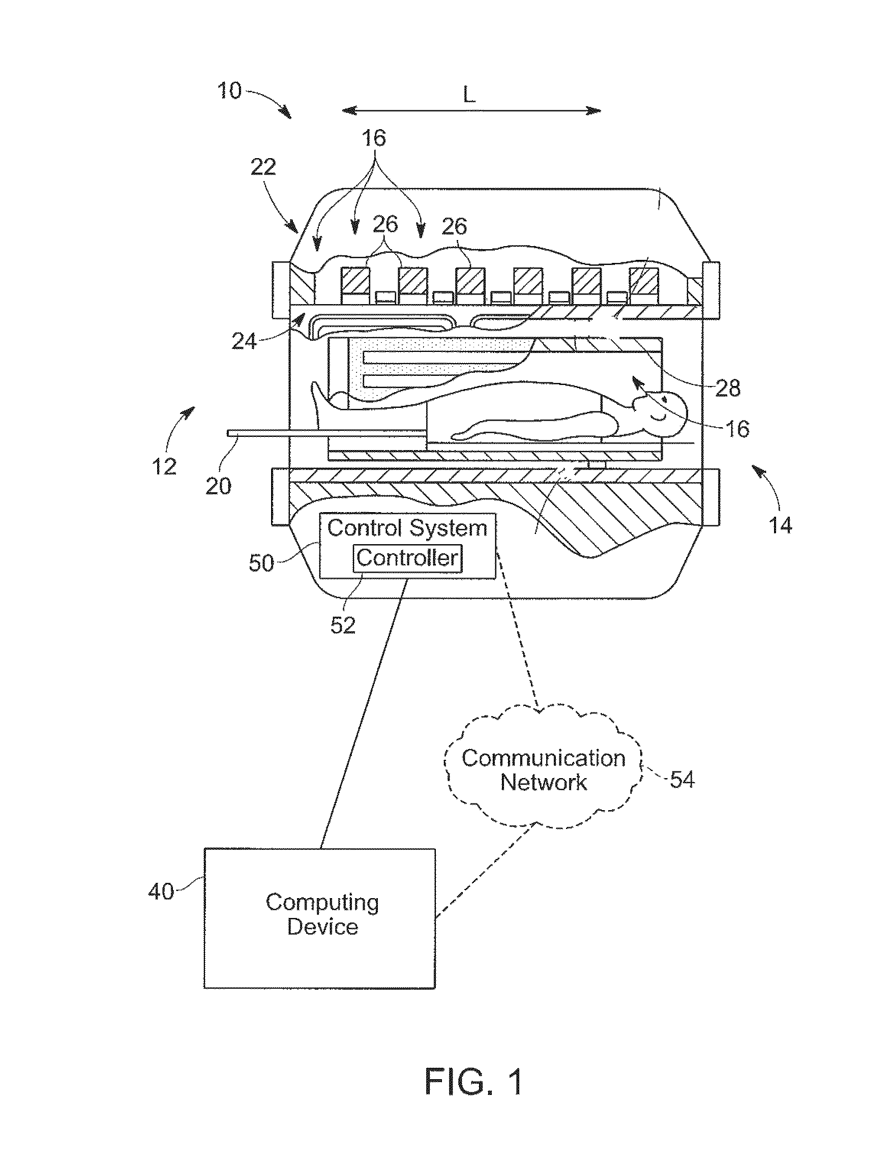
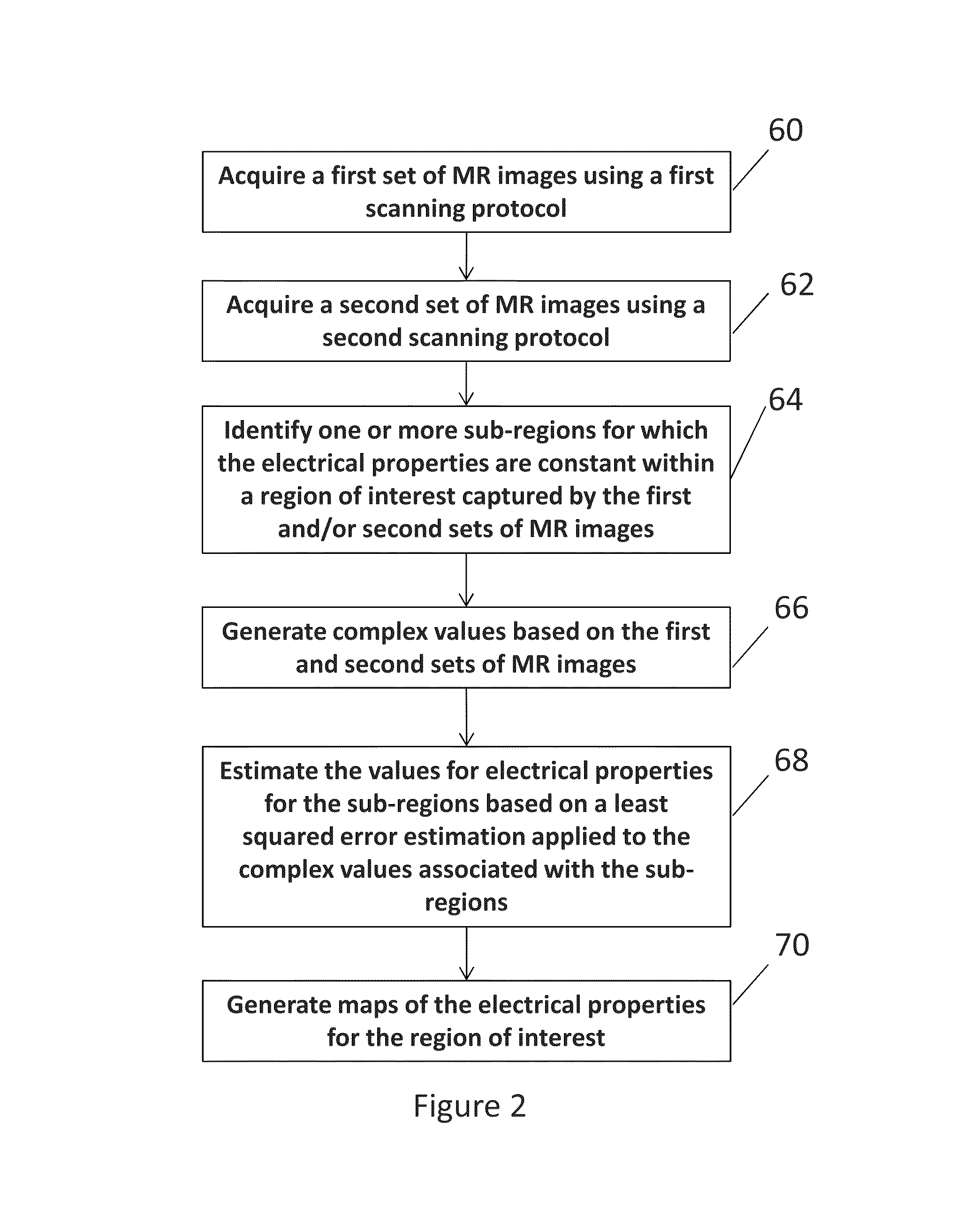
[0025]Exemplary embodiments provide a system and method to calculate the electrical properties of tissue (e.g., permittivity and / or conductivity) using a least squared error estimation based on a region of tissue having the same type (e.g., muscle, fat, bone). Exemplary embodiments of the present disclosure can allow for real time estimation of local Radio-Frequency (RF) power deposition or, in conjunction with mapping or images, provide diagnostically relevant information, such as for identifying tissue abnormality. At least one technical effect of some embodiments is the non-invasive estimation of the conductivity and / or permittivity of tissue using MRI in a clinically acceptable time frame. Other technical effects for some embodiments include evaluating RF safety, performing RF therapeutic methods, and diagnosing tissue abnormality using MRI mapping of conductivity and / or permittivity.

[0026]An intensity (magnitude) of pixels in an MR image of a region of interest can be used to i...


PUM


Abstract

Exemplary embodiments of the present disclosure are directed to estimating an electrical property of tissue using MR images. Complex values having real components and imaginary components are generated and are associated with pixels in one or more MR images that corresponding to a region of tissue for which the electrical property is constant. An estimated value of the electrical property for the region of tissue is determined based on a least squared error estimation applied to the complex values.

Description

STATEMENT AS TO FEDERALLY SPONSORED RESEARCH & DEVELOPMENT[0001]This invention was made with Government support under contract number R01CA154433 awarded by the National Institutes of Health through the National Institute of Biomedical Imaging and Bioengineering. The Government has certain rights in the invention.BACKGROUND[0002]Magnetic Resonance Imaging (MRI) or Nuclear Magnetic Resonance (NMR) imaging generally provides spatial discrimination of resonant interactions between radio frequency (RF) waves on nuclei in a magnetic field. Specifically, MRI utilizes hydrogen nuclear spins of the water molecules in the human body, which are polarized by a strong, uniform, static magnetic field, commonly referred to as B0 or the main magnetic field. When a substance, such as human tissue, is subjected to the main magnetic field, the individual magnetic moments of the spins in the tissue attempt to align with the main magnetic field. When excited by an RF wave, the spins precess about the m...

Claims


Application Information

Patent Timeline
Patent Type & Authority Patents(United States)
IPC IPC(8): G06K9/00G06T11/00G06K9/34G06K9/32G06V10/25
CPCG06K9/00G06K9/3233G06K9/342G06T11/003A61B5/055G01R33/246G01R33/48A61B5/4869G06V10/25G06V10/267G06F18/00
Inventor BULUMULLA, SELAKA BANDARAHANCU, ILEANALEE, SEUNG-KYUN
Owner GENERAL ELECTRIC CO
Why Eureka
Try Eureka