
Method and system for establishing the shape of the occlusal access cavity in endodontic treatment

a technology for occlusal access and endodontic treatment, which is applied in the field of method and system for establishing the shape of the occlusal access cavity in endodontic treatment, can solve the problems of improper preparation, affecting the healing effect of endodontic instruments, and reducing the healing of retained endodontic instruments, so as to achieve minimal bending of endodontic instruments, easy preparation, and saving chair time

Active Publication Date: 2015-09-22
DENTSPLY IMPLANTS NV +1
View PDF7 Cites 6 Cited by
  • Summary
  • Abstract
  • Description
  • Claims
  • Application Information

AI Technical Summary

Benefits of technology

The present invention provides clinicians with unambiguous guidelines on how to optimally shape the access cavity in function of patient specific information. This helps them plan root canal treatment more efficiently and reduces the need for exploration and uncertainty. Additionally, the invention enables easy preparation of the access cavity to ensure straight line access and minimal bending of the endodontic instruments to avoid their breakage. Overall, the invention saves time and increases the success of root canal treatment.

Problems solved by technology

A well known problem during this type of approach is related to fracture of the instruments.
In cases with a periapical lesion, retained fragments of endodontic instruments have been reported to reduce healing.
One of the most common causes for instrument fracture is improper preparation of the occlusal cavity that provides access to the pulp chamber and the connected root canals.
Often the size of the cavity is undersized or improperly shaped.
This leads to improper bending of the instruments during removal of the pulp, ultimately resulting in instrument failure.
In addition, the improper preparation of the access cavity may result in missing a main root canal e.g. the fourth mesio-buccal canal in a top molar tooth.
Since the efficacy of the endodontic treatment is highly dependent on the extent to which the diseased / infected tissue is removed, missing a canal may eventually result in treatment failure.
Incomplete access increases the stress on the files and leads to breakage.
For the practitioner it is however very difficult to adequately prepare the access cavity because of limited visibility and because the location of the root canal orifices is mostly uncertain (in 3D) prior to the start of the treatment.
Without reliable information about the root canals, in particular their amount and the localization of their orifices in the floor of the pulp chamber, there is a large degree of likelihood that the access cavity will not be properly dimensioned.
Moreover, in some cases too much of the healthy occlusal tissue may be removed, potentially compromising an easy prosthetic restoration of the tooth.
While this method provides information about the root canals and helps by means of a guide to localize the root canals intra operatively, it does not mention or teach how to prepare and shape the access cavity to the pulp chamber in such a manner that straight line access is realized for each of the identified root canals, without removing excess material from the tooth's occlusal surface.
Hence, instrument fracture may still occur.
According to the current state of the art there is no way to optimize the shape of the access cavity in function of the prosthetic restoration (e.g. inlay, crown) of the tooth subsequent to the endodontic treatment.
The described method and system however does not discuss shaping or preparing the access cavity to the pulp chamber, e.g. in order to prevent instrument fracture or optimize the prosthetic restorative capabilities after the endodontic intervention.

Method used


Image

  • Method and system for establishing the shape of the occlusal access cavity in endodontic treatment
  • Method and system for establishing the shape of the occlusal access cavity in endodontic treatment
  • Method and system for establishing the shape of the occlusal access cavity in endodontic treatment

Examples


Embodiment Construction

[0032]The present invention will be described with respect to particular embodiments and with reference to certain drawings but the invention is not limited thereto but only by the claims.

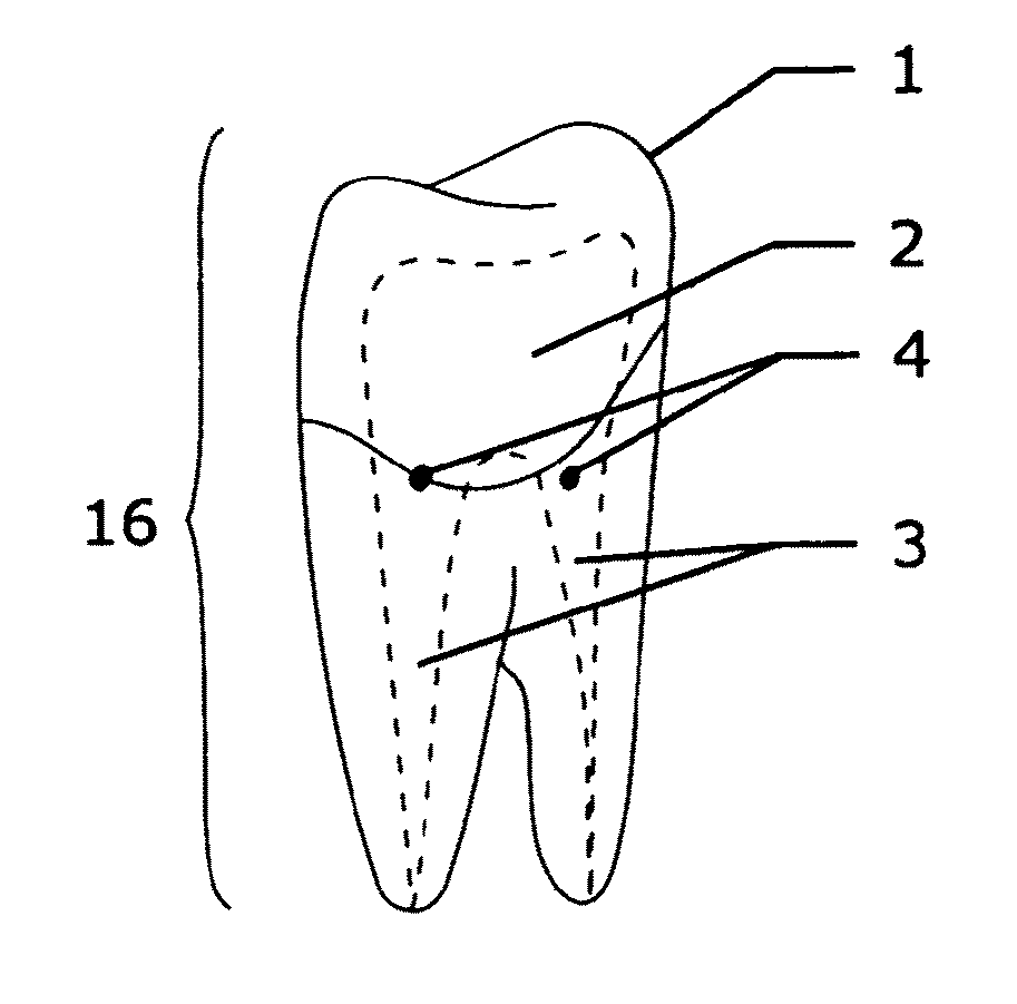
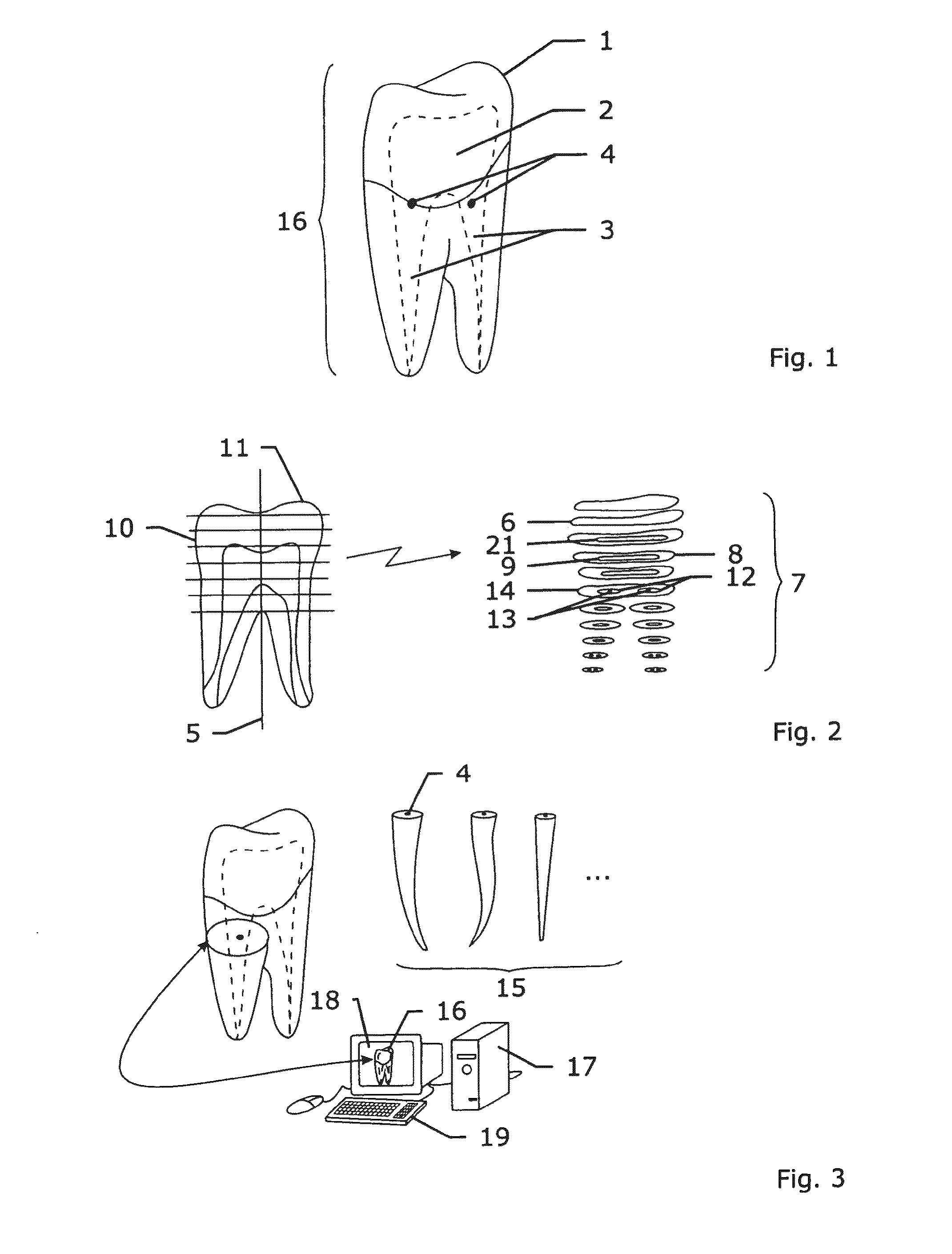
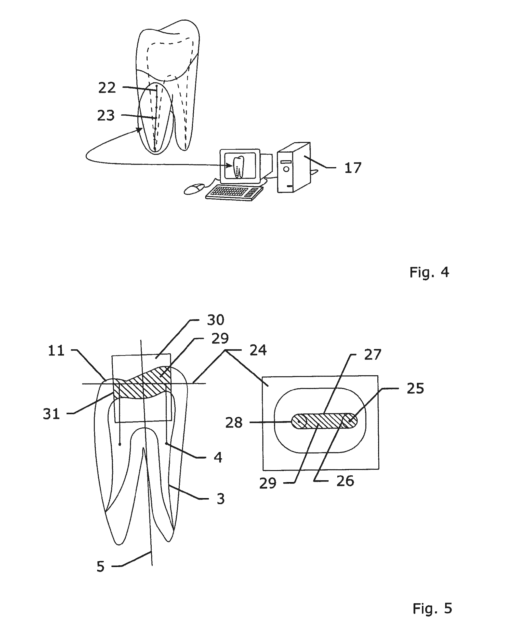
[0033]According to a preferred embodiment of the invention a three-dimensional computer model (16) of the tooth (1) including the pulp chamber (2) and the root canals (3), is required and obtained. Tooth enamel, bone and soft tissue are visible. The method starts when said three-dimensional computer model (16) is available. In a first subsequent step, the locations of the root canal orifices (4) (relative to the tooth's occlusal surface) are extracted from the computer model (16) of the tooth (1). According to one approach, this step is fully automated. As an example, the automation consists of the following sequence of actions performed by the system (17). First, the apical-coronal (longitudinal) axis (5) of the tooth (1) is determined. This can be done e.g. based on the calculation of the princip...


PUM


Abstract

A computer based method and system for defining and representing a shape and geometry of an occlusal access cavity to the tooth roots prior to endodontic treatment, include the step or elements for: loading onto the computer information of the geometry of a tooth obtained via one or more imaging techniques, creation of a 3D computer model of the tooth, including its internal architecture, visualization of the computer model, visualization of the location(s) of the entrance(s) to root canal(s) relative to the tooths occlusal surface, and based on the locations of the root canal orifices a shape of the access cavity is calculated.

Description

FIELD OF THE INVENTION[0001]The present application relates to methods, computer programs and software, tools and systems for establishing the shape of the occlusal access cavity in endodontic treatment.BACKGROUND[0002]During endodontic root canal treatment, the tooth pulp and nerves are removed from the root canals when they have been injured or are diseased. Treatment can be performed surgically by removing the apex of the tooth root (aka an apicoectomy) and sealing (obturating) the root canal via an access hole made through the jaw bone of the patient. However the more conventional treatment approach is to open the tooth occlusally, create a cavity (including the pulp chamber) providing access to the canal orifices, remove the diseased pulp and nerves, remove the diseased dentin by using specialized instruments such as endodontic files) to mechanically shape the root canals, clean and disinfect the root canals by means of specific irrigation media, and seal and obturate them to p...

Claims


Application Information

Patent Timeline
Patent Type & Authority Patents(United States)
IPC IPC(8): G06K9/00A61B19/00A61B5/055A61B6/03A61B8/14G06F19/12G06T19/20A61C5/02G06F19/00A61C13/00A61C5/40A61C5/42
CPCA61C5/023A61B5/055A61B6/032A61B8/14A61B19/50A61C5/02G06F19/12G06F19/3437G06T19/20A61B19/5244A61B2019/262A61B2019/505A61B2019/507A61C13/0004G06T2210/41A61B2090/502A61B34/20A61B34/10A61B2034/105A61B2034/107A61C5/40A61C5/42G16H50/50G16B5/00
Inventor VAN LIERDE, CARLPATTIJN, VEERLEVALLOTON, PAUL-HENRI
Owner DENTSPLY IMPLANTS NV
Learn More