A kind of injectable implant and preparation method thereof

A technology for injecting implants and implants, used in anti-inflammatory agents, pharmaceutical formulations, non-central analgesics, etc. Allergic reactions and other problems, to achieve good cell compatibility, good three-dimensional repair effect, good histocompatibility

Active Publication Date: 2017-09-08
SHAANXI RUISHENG BIOTECH
View PDF5 Cites 0 Cited by
  • Summary
  • Abstract
  • Description
  • Claims
  • Application Information

AI Technical Summary

Problems solved by technology

The disadvantages of the above bovine collagen implants are: a skin test is required 30 days before the injection to prevent allergies in the patient, the duration of the cosmetic repair effect is relatively short, and repeated injections are often required, and possible complications after the injection may cause Hemorrhagic spots, granulomas, acute allergies, delayed allergic reactions and systemic discomfort reactions, etc.
This patent only uses the physical milling method to prepare human hair keratin particle homogenate with a particle size of 60-80 μm. According to the inventor's experimental experience, different solid substances that are insoluble in water are physically milled without sieving, homogenization, etc. In the subsequent steps, the obtained particle size distribution range is usually 100-1000 μm, and it is difficult to obtain a smaller particle size range
[0017] Chinese patent CN02156797.2 discloses a preparation method of injectable collagen and its products and applications. In this patent, injectable collagen is prepared by extracting collagen fibers in animal tissues, and the original animal tissue will be completely destroyed during the preparation process. There are structural and other components, and the natural components with biological activity or biological function in natural tissues are lost, and it can only play a pure collagen filling role
[0029] At present, there is no product that uses amniotic membrane as raw material to prepare injectable filling materials, and there are no patents and literature reports

Method used


Image

  • A kind of injectable implant and preparation method thereof
  • A kind of injectable implant and preparation method thereof
  • A kind of injectable implant and preparation method thereof

Examples


Embodiment 1

[0074] Step 1. Disinfection treatment: Under aseptic conditions, soak the amniotic membrane in PBS until it is free of blood color and dirt, then wash it once with purified water, and then disinfect the amniotic membrane with 75% ethanol aqueous solution for 30 minutes minute;

[0075] Step 2, decellularization treatment: wash the amniotic membrane sterilized in step 1 with purified water for 3 times, then oscillate the amniotic membrane with 0.05mol / L sodium hydroxide aqueous solution for 30 minutes, the oscillation frequency is 50rpm, and finally use purified water Wash 3 times;

[0076] Step 3, modification treatment: the amniotic membrane obtained in step 2 was treated with 0.1 mol / L 1-ethyl-(3-dimethylaminopropyl)carbodiimide hydrochloride in an aqueous solution with a pH of 5 at 2° C. Under treatment for 6 hours;

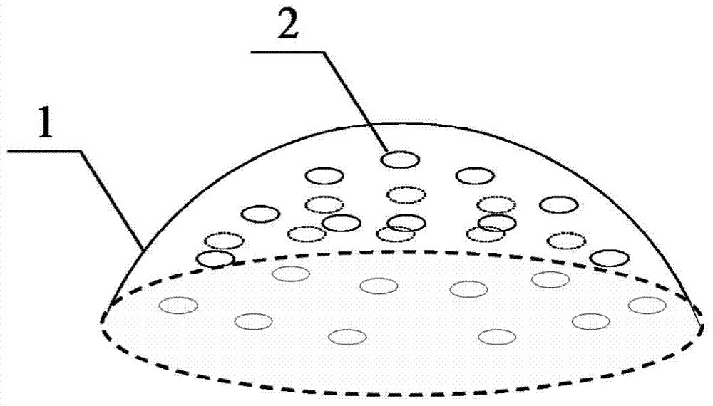
[0077] Step 4. Preparation of amniotic membrane microparticles: Wash the amniotic membrane modified in step 3 with purified water for 3 times, then vacuum f...

Embodiment 2

[0081] Step 1. Disinfection treatment: Under aseptic conditions, soak the amniotic membrane in PBS until it is free of blood color and dirt, then wash it with purified water for 5 times, and then disinfect the amniotic membrane with 75% ethanol aqueous solution for 15 minutes , and then disinfect the amnion with 0.1% peracetic acid for 15 minutes;

[0082] Step 2, decellularization treatment: wash the amnion after the disinfection treatment in step 1 with purified water once, and then use 0.01mol / L sodium hydroxide aqueous solution to shake the amnion for 10 minutes at a frequency of 80rpm, and then use 3mol / L sodium hydroxide solution to shake the amnion for 10 minutes. 1. L sodium chloride aqueous solution was shaken for 100 minutes, and finally cleaned 10 times with purified water;

[0083] Step 3, modification treatment: the amniotic membrane obtained in step 2 was treated with 0.5 mg / mL ribose aqueous solution at 2°C for 3 days;

[0084] Step 4. Preparation of amniotic m...

Embodiment 3

[0088] Step 1. Disinfection treatment: Under aseptic conditions, soak the amniotic membrane in PBS until it is free of blood color and dirt, then wash it with purified water for 3 times, and then disinfect the amniotic membrane with 75% ethanol aqueous solution for 10 minutes ; Disinfect the amniotic membrane with 1% hydrogen peroxide solution for 10 minutes;

[0089] Step 2, decellularization treatment: first wash the sterilized amniotic membrane twice with purified water, then oscillate the amniotic membrane with 1 mg / mL EDTA aqueous solution for 60 minutes, and oscillate the 0.1 mg / mL trypsin aqueous solution for 10 minutes. Both are at 60rpm, and finally washed with purified water for 6 times;

[0090] Step 3, modification treatment: treating the amniotic membrane obtained in step 2 with an aqueous glutaraldehyde solution with a volume concentration of 1% at room temperature for 6 hours;

[0091]Step 4. Preparation of amniotic membrane microparticles: Wash the amniotic me...


PUM

PropertyMeasurementUnit
concentrationaaaaaaaaaa
particle sizeaaaaaaaaaa
diameteraaaaaaaaaa

Abstract

The invention relates to an injectable implant and a preparation method thereof. The implant is as follows: amniotic membrane microparticles are mixed in sodium chloride phosphate physiological solution with a pH of 7.0-7.5. The preparation method is as follows: through the steps of disinfection treatment, decellularization treatment, modification treatment and preparation of amnion microparticles, that is, amniotic membrane microparticles are used as component A and sodium phosphate chloride physiological solution as component B, according to the mass volume ratio of 20 :1~100:1 oscillating and mixing, and prepare amnion microparticle suspension with a concentration of 20~100mg / mL by high-speed microfluidic equipment, and the particle size of amnion microparticles is 50~200μm. The invention retains the natural three-dimensional structure of human amniotic membrane and a large number of natural active ingredients, reduces the immune rejection caused by the source of raw materials of cosmetic injection products after clinical use, and is suitable for injection from the deep layer of the subcutaneous dermis to the superficial layer of the skin Corrects moderate to severe wrinkles or folds with good cosmetic results.

Description

technical field [0001] The invention belongs to the technical field of medical cosmetology and plastic surgery, and in particular relates to an injectable implant derived from amniotic membrane biomaterials and capable of promoting autologous collagen regeneration and a preparation method thereof. Background technique [0002] Soft tissue fillers have been widely used in cosmetic surgery for a century to improve facial contours, correct wrinkles, fill in sunken scars, and fill volumes (such as lips), etc. Today, there is an increasing demand for soft tissue fillers. [0003] The ideal soft tissue filler should be cheap and easy to obtain, good biological inertness, biocompatibility and stability, non-toxic, non-carcinogenic, non-infectious, no acute or chronic inflammatory response, easy to inject and remove, and able to maintain Long-term or even permanent cosmetic restoration with short recovery times. But so far, there is still no such ideal injectable filler. [0004] ...

Claims


Application Information

Patent Timeline
Patent Type & Authority Patents(China)
IPC IPC(8): A61F2/02A61K38/39A61K9/10A61P27/02A61P17/02A61P29/00
Inventor 刘博文张宏波冉永峰
Owner SHAANXI RUISHENG BIOTECH
Why Eureka
Try Eureka