
Systems and methods for electrical stimulation of sphenopalatine ganglion and other branches of cranial nerves

a technology of cranial nerves and electrical stimulation, which is applied in the field of systems and methods for electrical stimulation of sphenopalatine ganglion and other branches of cranial nerves, can solve the problems of nasal mucosa sloughing, difficult to reliably perform pterygopalatine fossa injection, and only a brief respite from pain

Active Publication Date: 2015-11-03
ELECTROCORE
View PDF53 Cites 0 Cited by
  • Summary
  • Abstract
  • Description
  • Claims
  • Application Information

AI Technical Summary

Benefits of technology

The present invention relates to a method and device for treating various disorders, such as headaches, by stimulating a specific nerve in the patient using a non-invasive technique. The method involves advancing electrode leads through one or both nostrils to the nasopharyngeal mucosa posterior to the middle turbinate adjacent to the SPG. The device is then placed on the mucosal surface and secured using medical glue or surgical net. The technical effects of this invention are that it can be used non-invasively and without damaging the surrounding blood vessels and nervous tissue. These technical effects offer improved safety and efficiency for treating disorders that have been treated with anesthetic blockade of the SPG.

Problems solved by technology

Unfortunately, the SPG swabbing produces only a brief respite from pain, whether by using a cotton swab as originally described by SLUTER, or by means of a topical administration device [U.S. Pat. No. 8,231,588, entitled Methods for ameliorating pain and devices for delivering a medicament, to XIA].
In addition, injection into the pterygopalatine fossa is difficult to perform reliably due to considerable anatomical variability of the patients, with damage to the maxillary artery that lies next to the SPG being not uncommon.
Furthermore, the nasal mucosa may slough during needle insertion.
This is not a preferred method because ablation would destroy useful neurophysiological functions of the SPG, notwithstanding the pain that the SPG may cause.
Avoidance of the transnasal route has apparently been due to the fact that surgical access to the pterygopalatine fossa through the nose is considered difficult, because of the protected position of the fossa and because of its complex and somewhat variable neurovascular anatomy.
Avoidance of a transnasal route has also been due to structural and energizing limitations of prior electrical stimulators, which would make their introduction through the nose essentially impossible.

Method used


Image

  • Systems and methods for electrical stimulation of sphenopalatine ganglion and other branches of cranial nerves
  • Systems and methods for electrical stimulation of sphenopalatine ganglion and other branches of cranial nerves
  • Systems and methods for electrical stimulation of sphenopalatine ganglion and other branches of cranial nerves

Examples


first embodiment

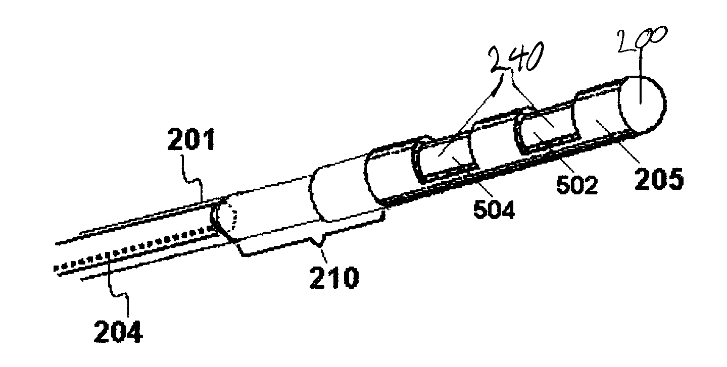
[0053]In the first embodiment, the stimulator is placed atop the mucosa area shown by hash marks in FIG. 6A, and medical glue such as cyanoacrylates or even denture adhesives are used to secure the stimulator to the mucosal surface. A surgical net may be placed over the stimulator to help keep it in place. Alternatively, the stimulator is attached to the underlying tissue using sutures that are placed through loops that are attached to the stimulator. This first, essentially noninvasive, embodiment might in some cases be considered to be temporary. When used temporarily, it may be used to determine whether the electrical stimulation will be effective, before using a more invasive approach. A significant advantage of this method is that there is no risk of damaging the blood vessels and nervous tissue within the pterygopalatine (sphenopalatine) fossa, because the fossa is not breached. Another advantage is its relative technical simplicity.

[0054]In the second embodiment (submucosal a...

third embodiment

[0055]The third embodiment is like the second, except that the anchor is inserted more permanently into a rigid structure such as the pterygoid process (PP in FIG. 6B) or the vomer. An example of such an anchor is shown in FIG. 7D. The stimulator is then attached to the anchor outside of the pterygopalatine (sphenopalatine) fossa at the site of attachment shown in FIG. 7D. Thus, the stimulator in the first three embodiments lies mostly outside the fossa.

fourth embodiment

[0056]In the fourth embodiment, a needle is inserted through the mucosa by approximately 1 mm, into the pterygopalatine (sphenopalatine) fossa, at a site that is unlikely to encounter blood vessels or nerves. The site may have been selected by using high resolution MR imaging of the contents of the fossa, ultrasound imaging with a transducer at the tip of a catheter inserted through the nostril, or by the statistical likelihood of a safe insertion at some site. A lead blank (e.g., teflon coated flexible wire, possibly with a spatula-shaped end) is then inserted into the lumen of the needle. The lead blank is advanced along a path of least resistance, e.g., through fat tissue, but being deflected from the more solid nerve and blood vessel tissue as it advances. Thus, the path that the lead blank takes is not necessarily known in advance, but instead opens a safe wormhole within the fossa. The lead blank is then withdrawn from the needle, and a catheter-like stimulator is advanced int...


PUM


Abstract

Devices, systems and methods are disclosed for modulating cranial nerves, such as the sphenopalatine ganglion, to treat a medical condition of a patient, such as cluster headache. A stimulation device is advanced transnasally to a target site at or adjacent to the nasopharyngeal mucosa posterior to the middle turbinate. Electrical impulses are applied through one or more electrodes in the stimulation device to the target nerve sufficient to modulate the nerve and treat the medical condition.

Description

CROSS REFERENCE TO RELATED APPLICATIONS[0001]The present application claims the benefit of U.S. Provisional Application Ser. No. 61 / 757,200 filed 27 Jan. 2013; which is incorporated herein by reference in its entirety.BACKGROUND OF THE INVENTION[0002]The field of the present invention relates to the delivery of energy impulses (and / or energy fields) to bodily tissues for therapeutic purposes. The invention relates more specifically to the use of electrical stimulation of the sphenopalatine ganglion (SPG) and other cranial nerves for treating disorders in a patient, such as primary headache.[0003]The sphenopalatine ganglion (SPG) is a nerve ganglion found in the pterygopalatine (sphenopalatine) fossa, close to the sphenopalatine foramen (102 in FIG. 3; also known as the pterygopalatine ganglion, ganglion pterygopalatinum, Meckel's ganglion, and nasal ganglion). There are two SPG, on either side of the nose. The SPG is usually triangular or oblong, and in humans it is approximately 5 ...

Claims


Application Information

Patent Timeline
Patent Type & Authority Patents(United States)
IPC IPC(8): A61N1/00A61N1/05A61N1/36A61N1/378A61N1/372
CPCA61N1/36075A61N1/0546A61N1/3787A61N1/37205A61N1/37229
Inventor SIMON, BRUCE J.ERRICO, JOSEPH P.
Owner ELECTROCORE
Learn More