Medical image processor and storage medium

a technology of medical image and storage media, applied in image enhancement, fluorescence/phosphorescence, instruments, etc., can solve the problems of poor prognosis of cases where her2 is positive, low accuracy, and high accuracy, and achieve the effect of efficient understanding of a cancer region

Active Publication Date: 2015-11-17
KONICA MINOLTA INC
View PDF19 Cites 6 Cited by
  • Summary
  • Abstract
  • Description
  • Claims
  • Application Information

AI Technical Summary

Benefits of technology

[0036]According to the present invention, it is possible to efficiently understand a cancer region where there is overexpression of a specific protein from the entire image of a tissue slice.

Problems solved by technology

HER2 protein is considered to be a prognostic factor of breast cancer, and it is known that especially in a case where lymph node metastasis is positive, the prognosis of cases where HER2 is positive is significantly poor.
Comparing the IHC method and the FISH method, the IHC method is easier, but there is the problem that accuracy is low.
Turning to the FISH method, the accuracy is high, but the process is troublesome, and the cost is high.

Method used


Image

  • Medical image processor and storage medium
  • Medical image processor and storage medium
  • Medical image processor and storage medium

Examples


Embodiment Construction

[0066]Embodiments of the present invention are described below with reference to the drawings, however, the present invention is not limited to the illustrated examples.

100>

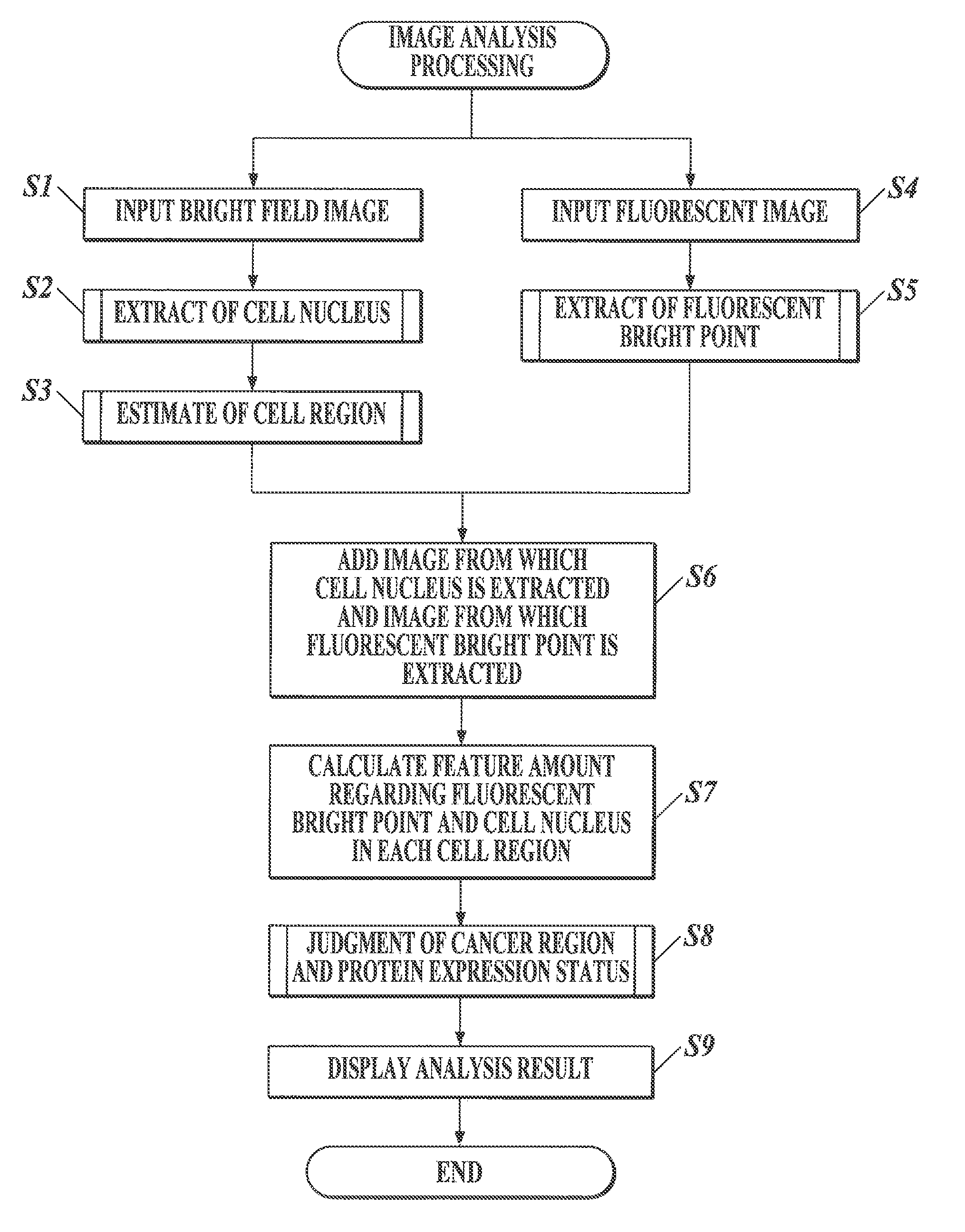
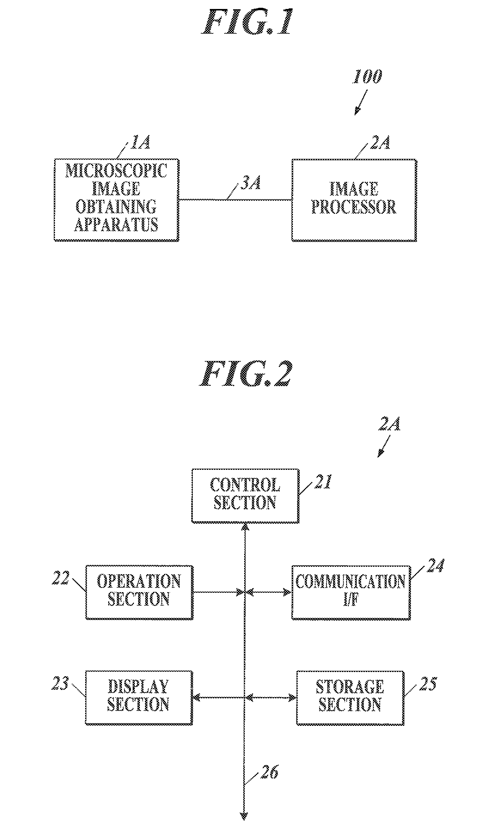
[0067]FIG. 1 shows an example of an entire configuration of a pathological diagnosis assistance system 100 of the first embodiment. The pathological diagnostic assistance system 100 obtains a microscopic image of a tissue slice of a human body stained with a predetermined staining reagent, and outputs a feature amount quantitatively expressing a specific biological substance in the tissue slice of the observation target by analyzing the obtained microscopic image.

[0068]As shown in FIG. 1, the pathological diagnosis assistance system 100 includes a microscopic image obtaining apparatus 1A and an image processor 2A connected to each other through an interface such as a cable 3A so as to be able to transmit and receive data. The method of connecting the microscopic image obtaining apparatus 1A and the image processo...


PUM

PropertyMeasurementUnit
thicknessaaaaaaaaaa
wavelengthaaaaaaaaaa

Abstract

A medical image processor and a storage medium are shown. According to one implementation, the medical image processor includes the following. An input unit is used to input a cell shape image and a fluorescent image showing expression of a specific protein. A cell nucleus extracting unit extracts a cell nucleus. A fluorescent bright point extracting unit extracts a fluorescent bright point. A region estimating unit sets a predetermined region. When the set region does not overlap with another, it is estimated to include one cell. When a plurality of the set regions overlap, it is estimated to include a plurality of cells. A feature amount calculating unit calculates a feature amount. A determining unit determines whether each estimated cell region is cancer and determines an expression status in the region based on the calculated feature amount. An output unit outputs a determination result.

Description

CROSS REFERENCE TO RELATED APPLICATION[0001]This Application is a 371 of PCT / JP2013 / 058919 filed on Mar. 27, 2013 which, in turn, claimed the priority of Japanese Patent Application No. JP2012-078722 filed on Mar. 30, 2012 both applications are incorporated herein by reference.TECHNICAL FIELD[0002]The present invention relates to a medical image processor and a program.BACKGROUND ART[0003]In pathological diagnosis, first, after processing of dehydrating the obtained tissue so as to be fixed and blocking with paraffin, the above is cut into thin pieces with a thickness of 2 to 8 μm, the paraffin is removed, and the above is stained to observe with a microscope. The pathologist performs diagnosis based on morphologic information such as change in size and shape of a nucleus of a cell, change in pattern as a tissue, etc., and stain information in the microscopic image. Methods known as the tissue staining method used in pathological diagnosis include a conventional chromatic staining m...

Claims


Application Information

Patent Timeline
Patent Type & Authority Patents(United States)
IPC IPC(8): G06K9/00G06T7/00G01N21/64
CPCG06K9/0014G06T7/0012G01N21/6458G06T2207/10061G06T2207/10064G06T2207/20221G06T2207/30024G06T2207/30096G06V20/695G06V20/698
Inventor TSUNOMORI, AKINORIOKADA, HISATAKEGONDA, KOHSUKEOHUCHI, NORIAKIWATANABE, MIKA
Owner KONICA MINOLTA INC
Why Eureka
Try Eureka