
Pathological diagnosis assisting apparatus, pathological diagnosis assisting method and non-transitory computer readable medium storing pathological diagnosis assisting program

a pathological diagnosis and assisting apparatus technology, applied in the field of pathological diagnosis assisting apparatus, can solve the problems of insufficient accuracy of diagnostic assisting information provided by the method based on the area occupancy rate of collagenous fiber alone in the stained sample, and the inability to use elastic fiber as an index, so as to improve the accuracy of pathological diagnosis and high-accuracy pathological diagnosis

Active Publication Date: 2016-03-15
KEIO UNIV +1
View PDF9 Cites 0 Cited by
  • Summary
  • Abstract
  • Description
  • Claims
  • Application Information

AI Technical Summary

Benefits of technology

[0011]Accordingly, it is an object of the present invention, in consideration of such a finding, to provide highly accurate pathological diagnosis assisting information based on the occupancy rates of the collagenous fiber and the elastic fiber in the tissue serving as the standards in detection of tissue fibrosis in image processing of the image of the stained sample. In a preferred mode of the present invention, the stage of tissue fibrosis is determined by using a total of the occupancy rates of the collagenous fiber and the elastic fiber, such that accuracy of determination by the pathological diagnosis assisting apparatus is improved. In another preferable mode, further, the stage of tissue fibrosis is determined by using the quantity of the elastic fiber slowly accumulated in the fibrous septa together with a quantity of the collagenous fiber, such that the new index enabling screening for a generation time of the fibrotic tissue is provided.

Problems solved by technology

However, the diagnosis assisting information provided by a method based on the area occupancy rate of the collagenous fiber alone in the stained sample is not sufficiently accurate.
Moreover, since a quantity of the elastic fiber in the tissue mentioned in Non-Patent Documents 2, 3 is small in comparison to the collagenous fiber, the elastic fiber has not been used as an index for assisting pathological diagnosis using image processing of the image of the stained sample.

Method used


Image

  • Pathological diagnosis assisting apparatus, pathological diagnosis assisting method and non-transitory computer readable medium storing pathological diagnosis assisting program
  • Pathological diagnosis assisting apparatus, pathological diagnosis assisting method and non-transitory computer readable medium storing pathological diagnosis assisting program
  • Pathological diagnosis assisting apparatus, pathological diagnosis assisting method and non-transitory computer readable medium storing pathological diagnosis assisting program

Examples


Embodiment Construction

[0034]A pathological diagnosis assisting apparatus according to one embodiment of the present invention will be described with reference to the accompanying drawings. A pathological diagnosis assisting method and a non-transitory computer readable medium storing a pathological diagnosis assisting program according to the present invention will become clear from the description of the pathological diagnosis assisting apparatus according to the embodiment of the present invention.

[0035]A sample used in the present embodiment is stained with, for example, EVG (Elastica Van Gieson) stein. EVG stein may stain a collagenous fiber and an elastic fiber in a tissue. Adopting this staining method to a lesional tissue obtained in a liver biopsy enables identification of the collagenous fiber in a liver cell. According to the present embodiment, in staining the sample with EVG stain, the elastic fiber is stained purple (dark purple) with Weigert's resorcin-fuchsin stain, a nuclear is stained bl...


PUM


Abstract

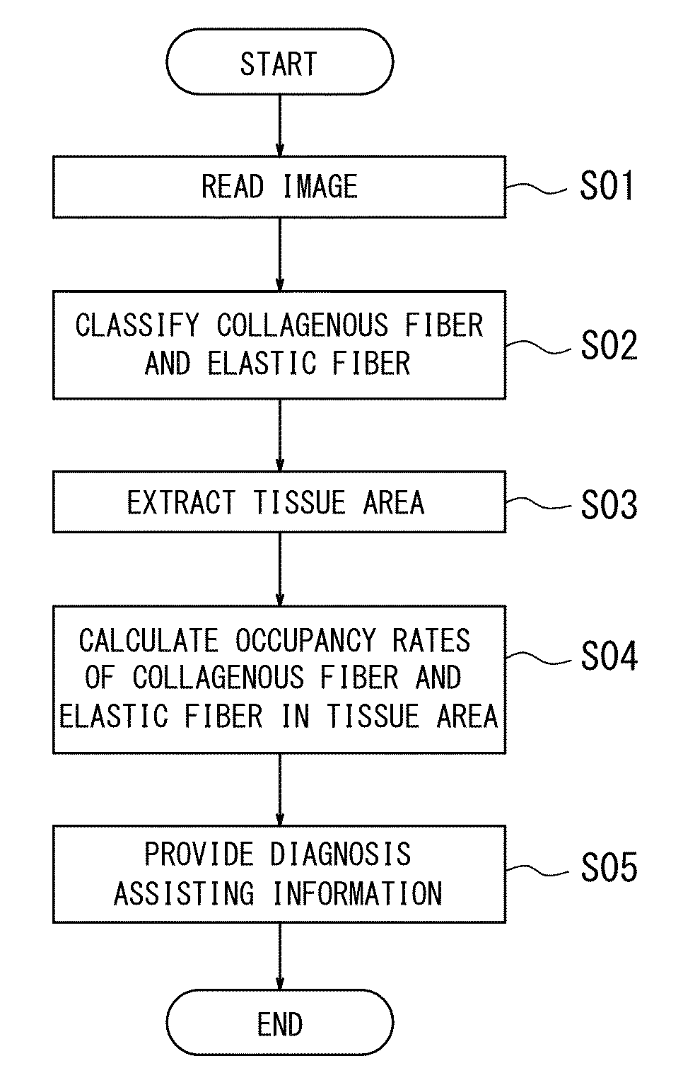
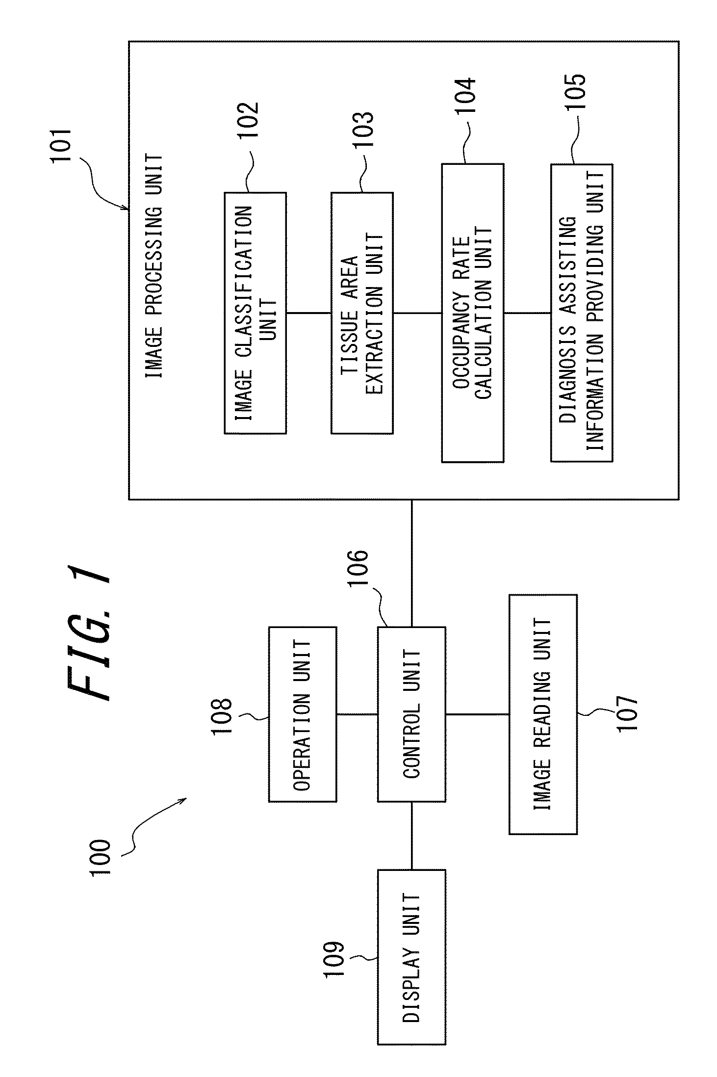
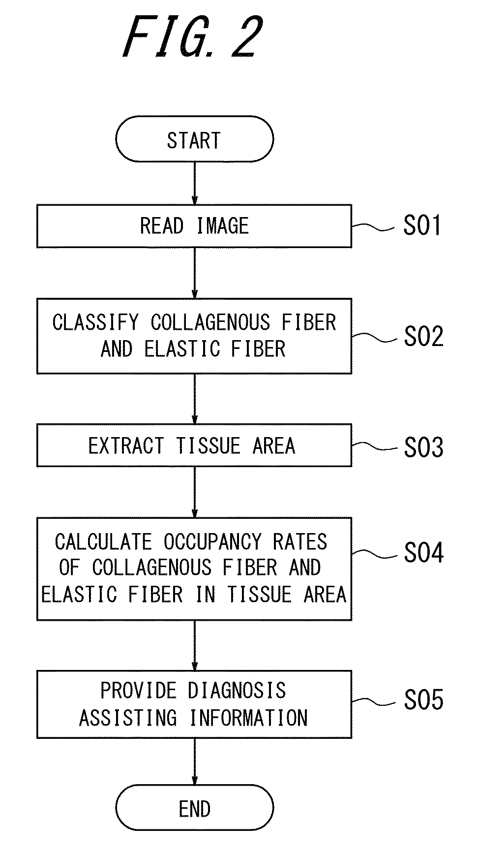
A pathological diagnosis assisting apparatus according to the present invention provides the diagnosis assisting information for assisting diagnosis of tissue fibrosis from the image of the stained sample, and includes an image reading unit configured to read the image of the stained sample and an image processing unit configured to process the image. The image processing unit includes an image classification unit configured to classify the collagenous fiber and the elastic fiber, a tissue area extraction unit configured to extract a tissue area, an occupancy rate calculation unit configured to calculate the occupancy rates of the collagenous fiber and the elastic fiber in the tissue area, and a diagnosis assisting information providing unit configured to provide the diagnosis assisting information based on the calculated occupancy rates of the collagenous fiber and the elastic fiber.

Description

FIELD OF THE INVENTION[0001]The present invention relates to a pathological diagnosis assisting apparatus, a pathological diagnosis assisting method and a non-transitory computer readable medium storing a pathological diagnosis assisting program.BACKGROUND OF THE INVENTION[0002]Progression of destruction and tissue fibrosis due to chronic inflammation in some organs are known to carry a risk of cancerogenesis. Assessment of a stage of tissue fibrosis is of great importance in order to find a phase of the progression of a lesion and, further, to determine the risk of the cancerogenesis.[0003]Chronic hepatitis is known as a disease which assessment of the stage of tissue fibrosis is of importance. As a method to assess the stage of tissue fibrosis, there is histopathological diagnosis. In histopathological diagnosis, a pathologist visually diagnoses a structural disorder due to fibrotic tissues. Generally, New Inuyama Classification is used for a diagnosis of the stage of fibrosis of ...

Claims


Application Information

Patent Timeline
Patent Type & Authority Patents(United States)
IPC IPC(8): G06K9/00G06T7/00
CPCG06T7/0012G06K9/00147G06T2207/10056G06T2207/20081G06T2207/30024G06V20/698
Inventor SAKAMOTO, MICHIIEHASHIGUCHI, AKINORIABE, TOKIYASAITO, AKIRASANO, MAKI
Owner KEIO UNIV
Learn More