

Assay system to identify therapeutic agents
a technology of assay system and therapeutic agent, applied in the field of assay system, to achieve the effect of facilitating the identification or qualification of pore formation, and reducing the binding of liposome reagen
- Summary
- Abstract
- Description
- Claims
- Application Information
AI Technical Summary
Benefits of technology
Problems solved by technology
Method used
Image

Examples
example 1
Two Color Vesicle Release Assay
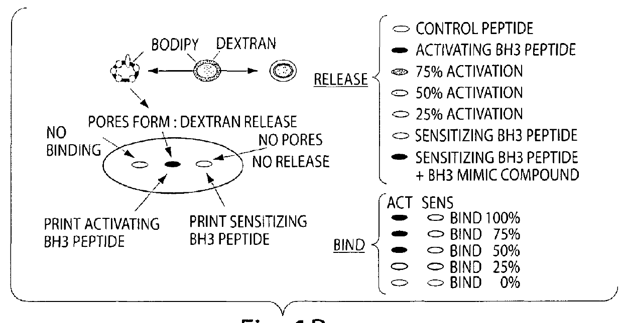
[0072]A simplified cell free system is used to detect Bax pore forming function on a lipid bilayer. The liposome release assay measures both the release of the fluorescently labeled contents and the lipid itself so a ratio of signals from each label or a shift in FRET will give accurate measurement of pore forming activity. Both a ratio of signals from each label and a shift in FRET are tested.
[0073]To accomplish this assay, purified proteins are incubated with synthetic liposomes labeled with BODIPY (either Phosphotidyl Inositol or Di-oc) and containing entrapped fluorescien dextran or coumeran dextran. The enhancement of Bax pore forming activity by BH3 peptide or BH3 mimic compounds is assessed in a liquid phase assay. Both colors will be read as well as the FRET signal generated from energy transfer between the fluorescence in the lumen of the liposome (coumeran or fluorescence) and that on the inner leaflet of the liposome. Efficiency is determine...
example 2
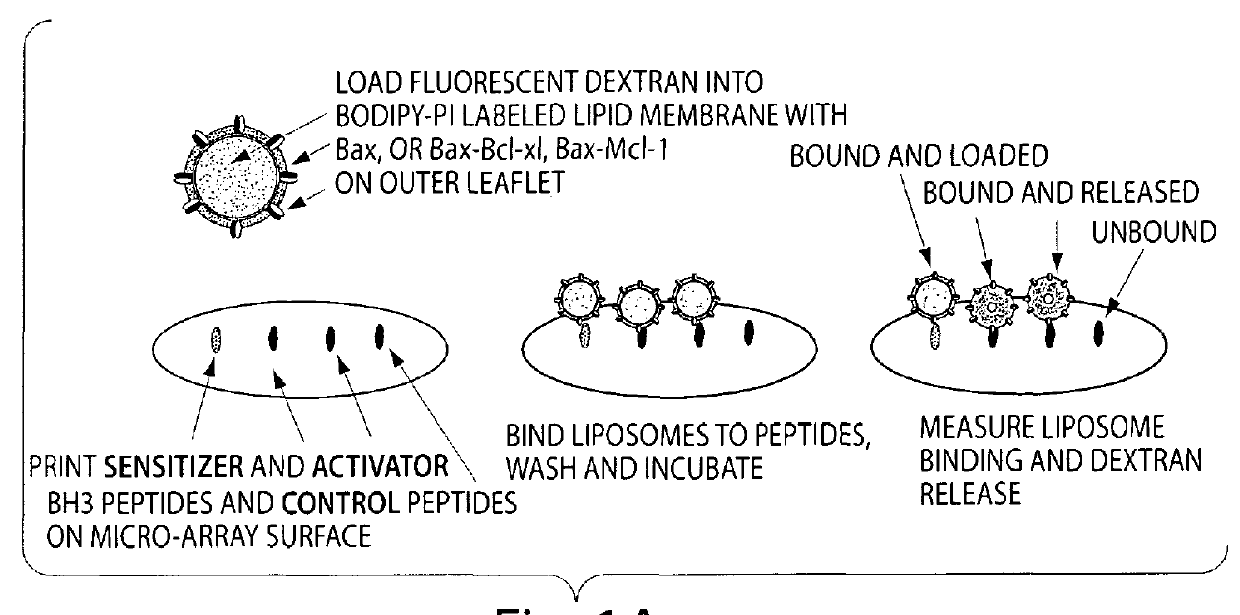
Assessment of Binding of the Two Color Lipid Vesicles to Printed BH3 Peptide Array
[0081]A peptide array format is used to assess the binding of Bax vesicles. These arrays are highly sensitive at detecting non-lipid associated Bcl-xL, Bcl-2 and Bax binding. Assays are prepared as described below, except that the format is changed to accommodate the traditional microscope slide. Using the microscope slide for a rough determination of a read out is sufficient for a first level experiment to identify that the system works. In a second level of experimentation, the format described above in Example 1 is used. Liposomes are prepared as described in Example 1, and incubated on the arrays and allowed to bind and incubated over a time course. Condition for optimal binding is determined by the liposome release profile and the readouts that indicate binding of molecules to the BH3 binding domains. Binding constants are calculated.
[0082]To generate the peptide arrays, peptides comprised of the ...
example 3
Optimization of Liposome Binding to Printed BH3 Peptides
[0083]Liposomes are labeled with combination of label determined in Example 1 above, and are prepared as described above. Liposome binding buffer includes PBS with other liposome compatible buffers. A phase 2 buffer is used without detergent, and the buffer specified in Kuwana et al can be used.
[0084]The Kds are established with purified proteins as a starting point and it is estimated that the amount of protein incorporated into a liposome is a direct function of the nature of the protein being used, and generally will agree with the amounts of protein incorporated into liposome for pore forming assays known in the art. The molar concentration of protein likewise follows generally that used for pore forming assays as described previously in the art.
[0085]Enough lipid is prepared to provide sufficient liposomes for the BH3 domain binding array, and is therefore loosely dependent on the amount of candidate therapeutic agents bei...
PUM
| Property | Measurement | Unit |
|---|---|---|
| diameter | aaaaa | aaaaa |
| diameter | aaaaa | aaaaa |
| number average sizes | aaaaa | aaaaa |
Abstract
Description
Claims
Application Information