Optogenetic probes for measuring membrane potential

a technology of optogenetic probes and membrane potentials, applied in the direction of instruments, peptides, fluorescence/phosphorescence, etc., can solve the problems of slow electrophysiological experiment setup, inability to access deep buried tissues, and difficult to measure membrane potentials, etc., to improve sensitivity, improve properties, and increase brightness

Active Publication Date: 2016-12-13
PRESIDENT & FELLOWS OF HARVARD COLLEGE +1
View PDF54 Cites 8 Cited by
  • Summary
  • Abstract
  • Description
  • Claims
  • Application Information

AI Technical Summary

Benefits of technology

[0009]By optically measuring the membrane potential of cells and sub-cellular compartments, the inventive polypeptides are capable of indicating electrical dynamics with sub-millisecond temporal resolution and sub-micron spatial resolution. The inventive polypeptides have improved properties over the wild-type Archaerhodopsin such as increased brightness, increased sensitivity, higher signal-to-noise ratios, increased linearity with respect to voltage or intensity, and faster response time (increased time resolution), with speed and sensitivity being important parameters for evaluating voltage indicators. The improved polypeptides provided herein are useful as optically detectable sensors for sensing voltage across membranous structures. It was previously demonstrated that the membrane potential in a membrane containing Archaerhodopsin 3 (Arch 3) can alter the optical properties of the protein, thereby making Arch 3 a voltage sensor. The modified microbial rhodopsin, Arch 3 D95N, has a 40 ms response time and lacks photoinduced proton pumping. Although the slower response time of this construct hampers detection of membrane potential and changes thereto in neurons, the Arch 3 D95N is fast enough to indicate membrane potential and action potentials in other types of cells, for example, in cardiomyocytes and does not perturb membrane potential in the cells wherein it is used.
[0010]Through a combination of directed evolution and targeted mutagenesis, polypeptides based on the human codon-optimized sequence of Achaerhodopsin genetically encoded voltage indicators GEVIs with improved performance have been identified.
[0026]In nature, microbial rhodopsins contain a bound molecule of retinal which serves as the optically active element. These proteins will also bind and fold around many other chromophores with similar structure, and possibly preferable optical properties. Analogues of retinal with locked rings cannot undergo trans-cis isomerization, and therefore have higher fluorescence quantum yields (Brack et al. Biophys. J. 65, 964-972 (1993)). Analogues of retinal with electron-withdrawing substituents have a Schiff base with a lower pKa than natural retinal and therefore may be more sensitive to voltage (Sheves et al. Proc. Nat. Acad. Sci. U.S.A. 83, 3262-3266 (1986); Rousso, I., et al. Biochemistry 34, 12059-12065 (1995)). Covalent modifications to the retinal molecule may lead to voltage-indicating proteins (VIPs) with significantly improved optical properties and sensitivity to voltage.
[0043]As used herein, the term “targeting sequence” refers to a moiety or sequence that homes to or preferentially associates or binds to a particular tissue, cell type, receptor, organelle, or other area of interest. The addition of a targeting sequence to an optical sensor composition will enhance the delivery of the composition to a desired cell type or subcellular location. The addition to, or expression of, a targeting sequence with the optical sensor in a cell enhances the localization of the optical sensor to a desired location within an animal or subject.
[0044]As used herein, the phrase “homologous mutation in another microbial rhodopsin that corresponds to the amino acid mutation in bacteriorhodopsin” refers to mutation of a residue in a desired microbial rhodopsin that is expected to have a similar effect to a substantially similar mutation in bacteriorhodopsin. One of skill in the art can easily locate a homologous residue in their desired microbial rhodopsin by performing an alignment of conserved regions of the desired microbial rhodopsin with a bacteriorhodopsin sequence using a computer program such as ClustalW. Examples of homologous mutations include the mutations made in the Examples set forth in this application.

Problems solved by technology

In spite of its fundamental biological role, membrane potential is very difficult to measure.
Electrophysiological experiments are slow to set up, can only be performed on one or a few cells at a time, cannot access deeply buried tissues (e.g., in vivo), do not work for cells that are too small (e.g. bacteria) or are enclosed in a hard cell wall (e.g. yeast), or are motile (e.g., sperm), cannot be applied to long-term measurements, and usually damage or kill the cell under study.
Simultaneous optical stimulation and readout of neural activity have been implemented via several combinations of the above techniques.17-21 However, robust genetically targeted all-optical electrophysiology has not been achieved due to limitations on the speed and sensitivity of genetically encoded voltage indicators (GEVIs), and spectral overlap between existing GEVIs and optogenetic actuators.

Method used


Image

  • Optogenetic probes for measuring membrane potential
  • Optogenetic probes for measuring membrane potential
  • Optogenetic probes for measuring membrane potential

Examples


example 1

Directed Evolution and Engineering of an Arch-Based Voltage Indicator

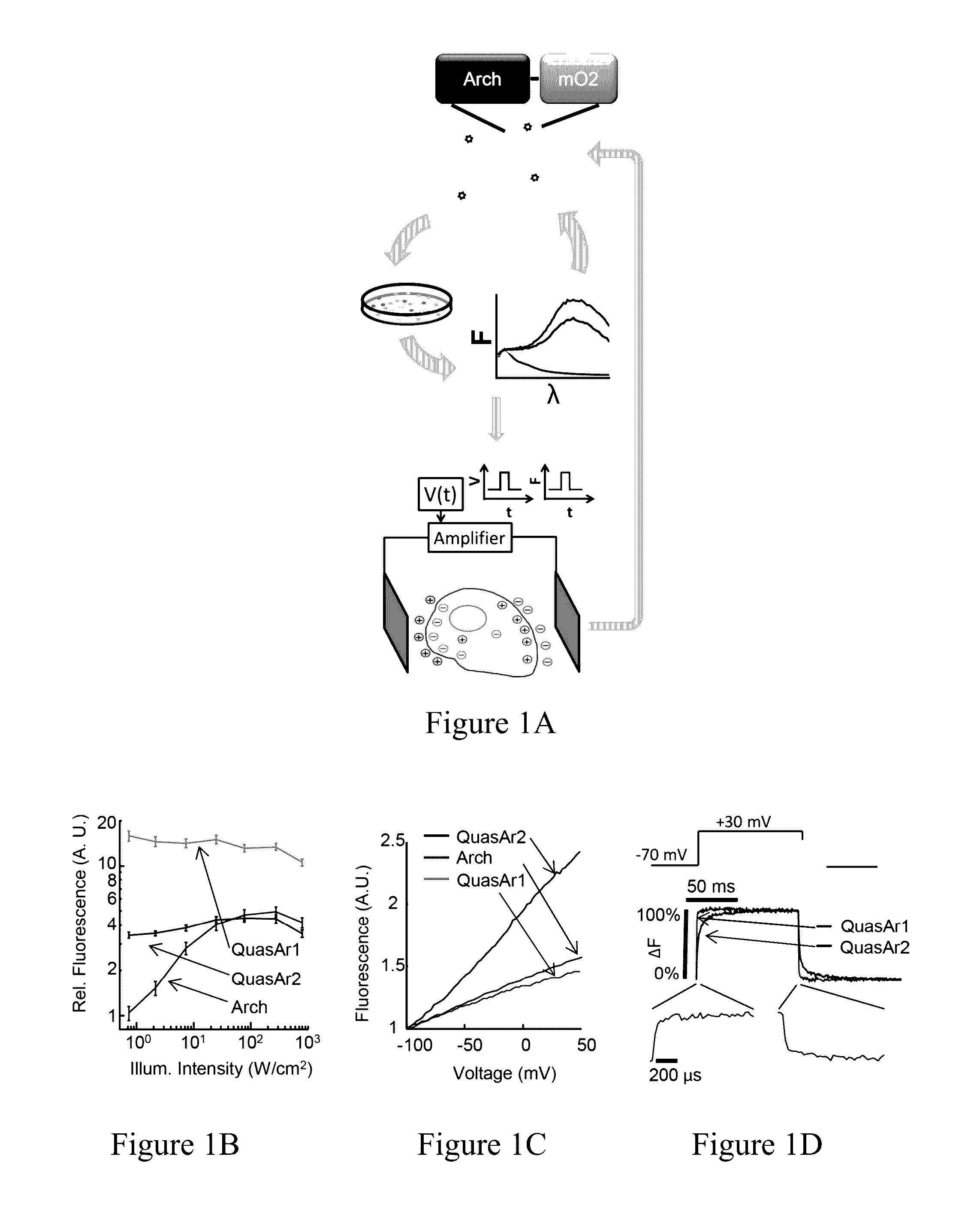
[0399]We previously showed that Archaerhodopsin 3 (Arch) functions as a fast and sensitive voltage indicator.10 Arch has the furthest red-shifted spectrum of any GEVI, giving it the unique property of little spectral overlap with channel rhodopsin actuators and GFP-based reporters. Thus it is natural to pair Arch-based indicators with optogenetic actuators for crosstalk-free all-optical electrophysiology.

[0400]However, wild-type Arch had some undesirable attributes for a reporter: it was very dim, and the brightness was a nonlinear function of illumination intensity.24 Illumination for imaging generated a hyperpolarizing photocurrent, which partially suppressed neural firing. The mutant Arch(D95N) did not pump, but its step response was dominated by a 41 ms time constant, too slow to resolve action potential (AP) waveforms.

[0401]We sought to repair these defects in engineered mutants of Arch. To accommodate the mul...

example 2

Comparison of QuasArs to Firelight

[0412]We compared the QuasArs to Arclight A242, a recently introduced GFP-based GEVI.23 Photophysical comparisons were performed in HEK cells, and action potential comparisons were performed in matched neuronal cultures (Methods). Arclight showed voltage sensitivity of −32±3% ΔF / F per 100 mV (n=7 cells; FIG. 9), comparable in magnitude to QuasAr1 and 2.8-fold smaller than QuasAr2. Arclight showed hi-exponential kinetics in response to rising or falling voltage steps (FIG. 9, Table 6). Mean half-response times were 42±8 ms and 76±5 ms on rising and falling edges at 23° C. (n=6 cells) and 11±1 and 17±2 ms on rising and falling edges at 34° C. (n=7 cells). Under continuous illumination at standard imaging intensity (488 nm, 10 W / cm2)11 Arclight photobleached with a time constant of 70 s. In cultured neurons, Arclight reported action potentials with an amplitude of ΔF / F=−2.7±0.5% (n=5 cells) and a single-trial signal-to-noise ratio (SNR) of 8.8±1.6 when...

example 3

Electrochromic Fluorescence Resonance Energy Transfer (eFRET)-Based GEVIs

[0414]Importance of Spectral Tunability in GEVIs.

[0415]The spectral range of GEVIs is important when combining GEVIs with other GEVIs, other optical reporters, or optogenetic actuators. Having GEVIs of multiple colors enables multiplex voltage imaging when distinct structures cannot be spatially resolved. For instance, to study separately excitatory and inhibitory neurons in intact tissue would require two colors of GEVIs. Furthermore, GFP-based GEVIs cannot be paired with other GFP-based reporters, e.g. gCaMP reporters of Ca2+,9 iGluSnFR reporter of glutamate,80 Perceval reporter of ATP,81 Clomelion reporter of Cl−,82 Pyronic reporter of pyruvate.83 GFP-based reporters also experience severe optical crosstalk with all optogenetic actuators. Even the reddest channel rhodopsin variants retain ˜20% activation with blue light used for GFP excitation.84

[0416]Spectral range is also important for imaging in intact t...


PUM

PropertyMeasurementUnit
response timeaaaaaaaaaa
voltageaaaaaaaaaa
voltageaaaaaaaaaa

Abstract

Provided herein are variants of an archaerhodopsin useful for application such as optical measurement of membrane potential. The present invention also relates to polynucleotides encoding the variants; nucleic acid constructs, vectors, cells comprising the polynucleotides, and cells comprising the polypeptides; and methods of using the variants.

Description

RELATED APPLICATIONS[0001]The present application claims priority under 35 U.S.C. §119(e) to U.S. provisional patent application, U.S. Ser. No. 62 / 013,775, filed Jun. 18, 2014, which is incorporated herein by reference.GOVERNMENT SUPPORT[0002]This invention was made with government support under grant numbers 1-R01-EB012498-01 and 1DP2OD007428 awarded by the National Institutes of Health (NIH), and under grant number N00014-11-1-0549 awarded by the Office of Naval Research (ONR). The government has certain rights in the invention.BACKGROUND[0003]Membrane-enclosed biological structures can support a voltage difference between the inside and the outside of the membrane. This voltage, also called a membrane potential, serves a variety of biological functions, including carrying information (e.g., in neurons), acting as an intermediate in the production of ATP (e.g., in bacteria and mitochondria), powering the flagellar motor (e.g., in bacteria), and controlling the transport of nutrien...

Claims


Application Information

Patent Timeline
Patent Type & Authority Patents(United States)
IPC IPC(8): G01N21/64C07K14/215C07K14/705G01N33/566
CPCC07K14/705C07K14/215G01N21/6428G01N33/566G01N2021/6441G01N2333/726
Inventor COHEN, ADAM E.HOCHBAUM, DANIELZOU, PENGFARHI, SAMOUIL LEONCAMPBELL, ROBERT EARLZHAO, YONGXINHARRISON, DANIEL JED
Owner PRESIDENT & FELLOWS OF HARVARD COLLEGE
Why Eureka
Try Eureka