
Method for the spectral identification of microorganisms

a microorganism and spectral data technology, applied in the field of microorganisms identification using spectral data, can solve the problems of waning ir bacteria identification, time-consuming and impractical procedures, and inability to use ir spectroscopy to identify bacteria as useful techniques, etc., to achieve rapid and reliable identification.

Inactive Publication Date: 2017-01-24
MCGILL UNIV +1
View PDF3 Cites 3 Cited by
  • Summary
  • Abstract
  • Description
  • Claims
  • Application Information

AI Technical Summary

Benefits of technology

The patent describes a way to quickly and reliably identify unknown microbes for medical diagnosis, food and environment control, and other purposes.

Problems solved by technology

However, it was concluded at that time that, although individual strains of bacteria definitely exhibit unique IR spectra, the identification of bacteria by IR spectroscopy could not be regarded as a useful technique, as the procedure was too time-consuming and impractical.
Thus, the above requirement imposes stringent conditions on spectral reproducibility, and interest in IR bacteria identification waned in the 1960s largely because these conditions could not be achieved with the IR instrumentation available at the time
However, such analyses have yielded relatively poor reproducibility and identification success rate.
Both these steps represent bottlenecks that have prevented the speed advantages of FTIR bacteria identification from being fully exploited.
Furthermore, they make building a spectral library a laborious and time consuming process, which likely accounts, in part, for the lack of commercial bacterial infrared spectral databases that would be required in order for FTIR bacteria identification to be implemented in routine microbiological analysis.
However, when microorganisms are deposited on a surface or suspended in a solution for acquisition of spectral data, there usually results an inhomogeneous distribution of the microorganisms within the sample.
Conventional acquisition of spectral data does not discriminate regions of inhomogeneities and therefore provides an “average” signal, which may comprise signal contributions that render the spectral information less reliable for identifying microorganisms.

Method used


Image

  • Method for the spectral identification of microorganisms
  • Method for the spectral identification of microorganisms
  • Method for the spectral identification of microorganisms

Examples


example 1

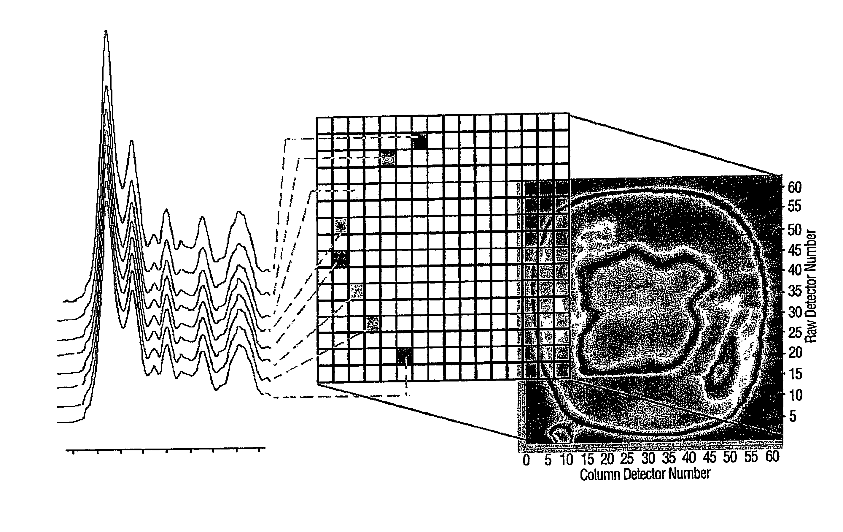
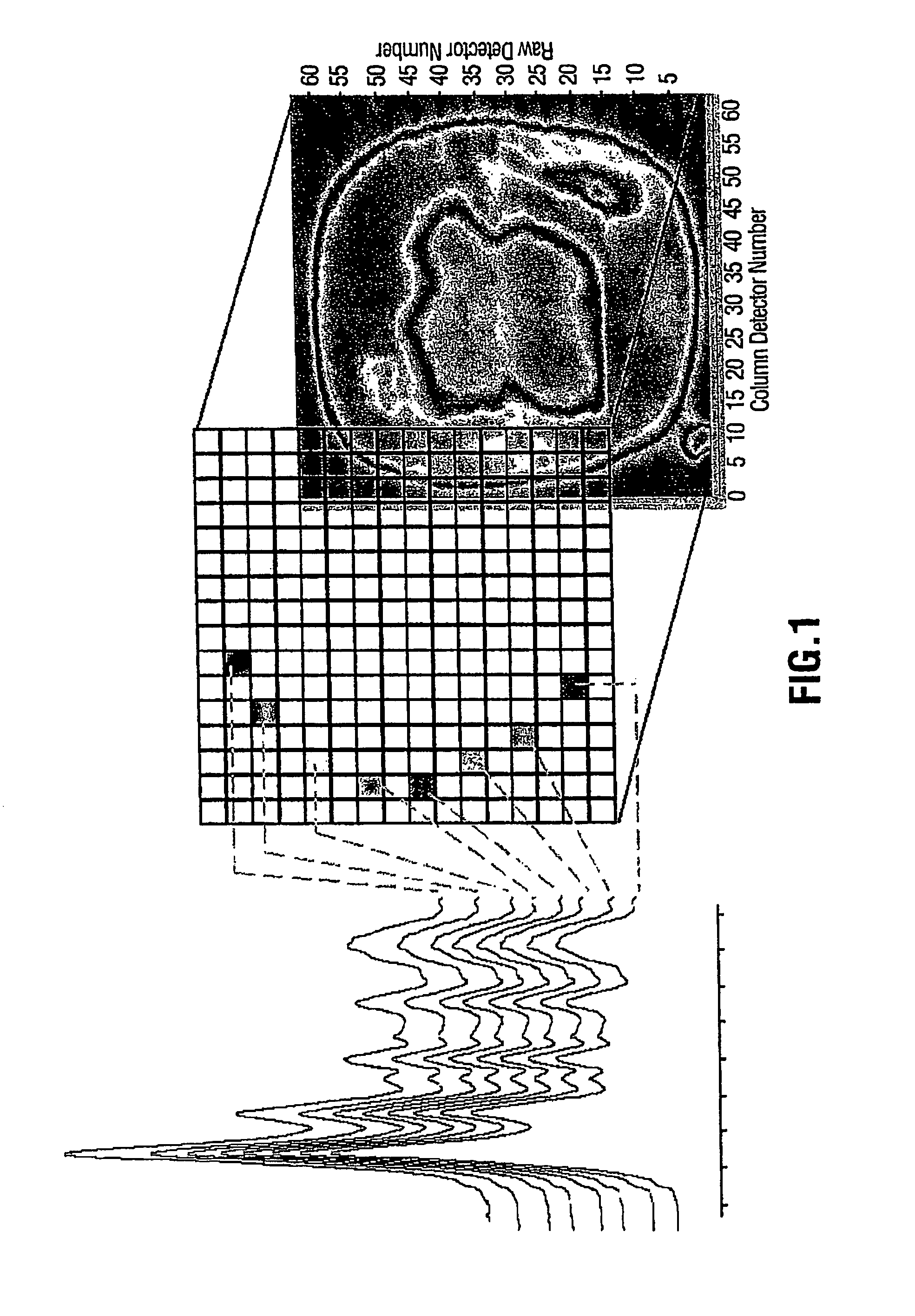
[0045]In this experiment 80 bacterial samples were characterized by FPA-FTIR. All spectra were collected on a Digilab FastIR imaging spectrometer equipped with a Digilab UMA-600 infrared microscope and a 16×16 MCT focal plane array detector (Digilab, Randolph, Mass., USA) operating under Win-IR Pro 3.3 (Digilab) by co-adding 256 scans at a resolution of 8 cm−1. In order to produce an absorbance, all single beam spectra and focal plane array imaging spectra were ratioed against the open beam spectrum of the sample. A constant flow of dry air was employed to purge the spectrometer and the microscope of carbon dioxide and water vapor. In total, (for the 80 bacterial samples) over 200,000 spectra of bacteria and bacterial cell extracts were recorded from the spectral images, yielding an average of 250 good pixels out of a possible 256 pixels per image, (each pixel generates a unique spectrum). A sterile wood toothpick was inserted into isolated colonies of bacteria to gently scrape some...

example 2

[0049]For this evaluation of FPA-FTIR spectroscopy, two Digilab Stingray systems, one equipped with a 16×16 and the other with a 32×32 array detector, were used.

Experimental Protocols

[0050]Growth of Bacteria.

[0051]Spectra of bacteria can be recorded from intact cells taken directly from culture plates. As early as the 1950s, it was recognized that the IR spectra of living bacterial cells strongly depend on the composition of the growth medium and time of growth. Consequently, extremely precise metabolic control and strict standardized handling of all samples is preferable to yield sufficient spectral reproducibility for comparison of the IR spectra of bacteria. To facilitate the use of FTIR spectroscopy in routine microbiological analysis, “Universal Medium” (UM™) was used. To date, this medium has supported the growth of virtually all bacteria and yeasts that have been tested. All 100 strain employed in the present work were grown on UM™ agar plates (provided by Quelab Laboratories...

example 3

Materials and Methods

Microbiological Specimens

[0062]A total of 8 strains of C. botulinum (CK2 A, 2 B, 17 B, 13983 B, Bennett E, Russ E, H461297 F, and 602 F) were used in this study. This investigation was only concerned with strains of C. botulinum in groups A, B, E, and F because only these serotypes give rise to intoxications in humans. The isolates were confirmed as C. botulinum because of the production of characteristic botulinum neurotoxins.

FTIR Spectroscopic Methods

Sample Preparation

[0063]Prior to FTIR spectral acquisition, all strains were incubated for 48 hours on both Brain Heart Infusion (BHI) and McClung Toabe with egg yolk (MTEYE) media. To optimize the growth conditions, proteolytic strains were grown at 35° C., while non-proteolytic strains were grown at 25° C. The bacteria were transferred to an infrared transmitting window (ZnSe). The bacteria were air dried on the IR window for 10 minutes and then incubated in formaldehyde vapours for several hours to ensure inact...


PUM

PropertyMeasurementUnit
spectrumaaaaaaaaaa
spectral intensityaaaaaaaaaa
chemical inhomogeneitiesaaaaaaaaaa

Abstract

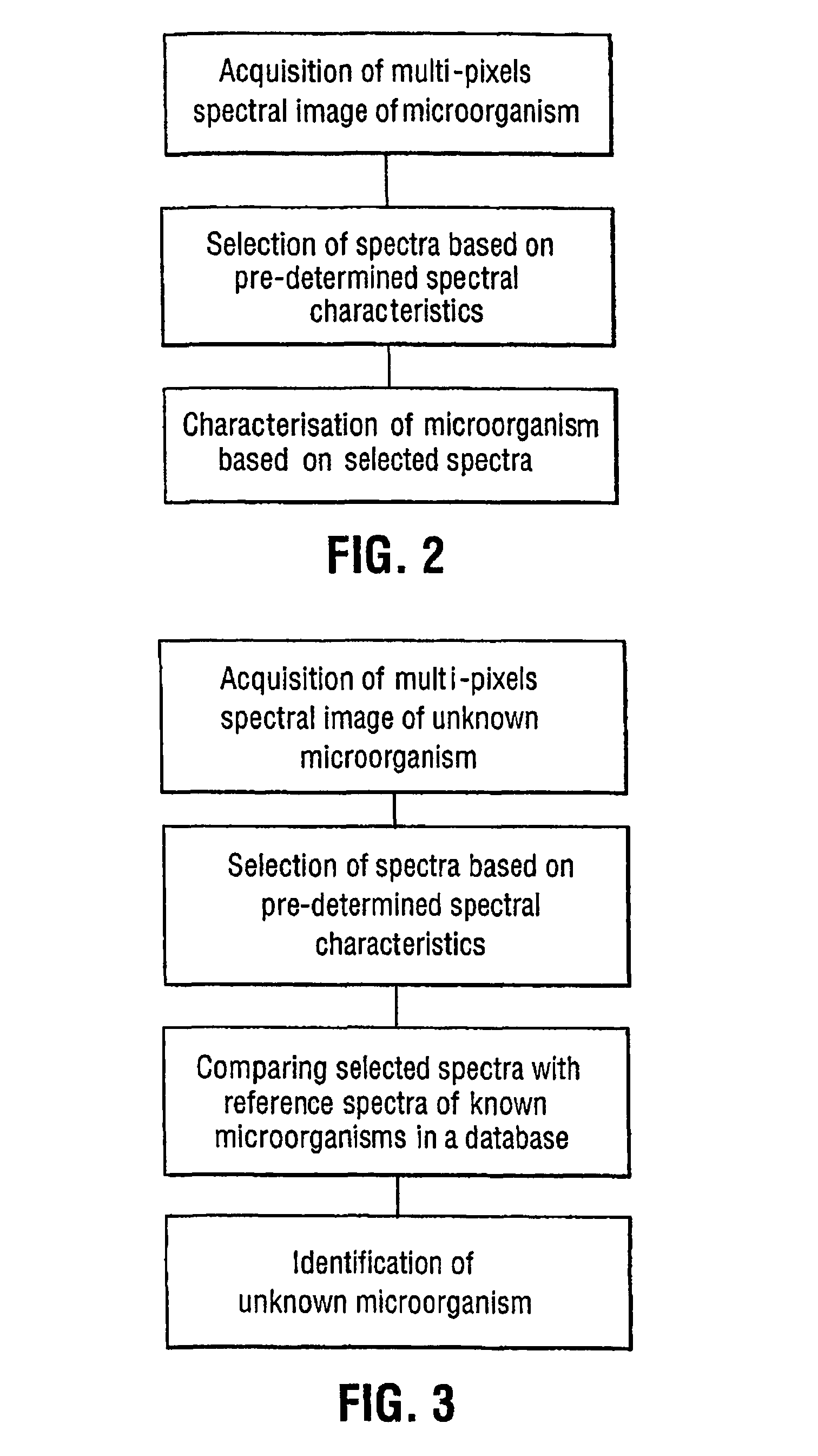
A rapid method for characterizing and identifying microorganisms using Focal-Plane Array (FPA)-Fourier Transform Infrared (FTIR) spectroscopy is disclosed. Multi-pixels spectral images of unknown microorganisms spectra are analyzed and compared to spectra of reference microorganisms in databases. The method allows rapid and highly reliable identification of unknown microorganisms for the purpose of medical diagnosis, food and environmental control.

Description

FIELD OF THE INVENTION[0001]The present invention relates to methods of microorganisms identification using spectral data.BACKGROUND OF THE INVENTION[0002]Fourier transform infrared (FTIR) spectroscopy is a known technique for the identification of microorganisms (Mariey et al., 2001 Vibr. Spectrosc. 26:151). Infrared (IR) spectroscopy measures the vibrations of chemical bonds within all the biochemical constituents of cells, i.e., proteins, lipids, polysaccharides, and nucleic acids, and provides quantitative information about the total biochemical composition of the intact whole microorganism. Furthermore, because the IR spectra of microorganisms consist of distinct and unique patterns the spectra effectively serve as “fingerprints,” allowing for their use in taxonomic discrimination. Indeed, the use of IR spectroscopy as a means of differentiating and identifying bacteria was extensively reported as early as the 1950s. However, it was concluded at that time that, although individ...

Claims


Application Information

Patent Timeline
Patent Type & Authority Patents(United States)
IPC IPC(8): G01N33/48G01N33/50G01N21/3563G06F19/12G06F19/18G01N21/17G01N21/35G16B5/00G16B20/00
CPCG01N21/3563G06F19/12G06F19/18G01N2021/1748G01N2021/1772G01N2021/3595G01N2201/129G16B5/00G16B20/00
Inventor ISMAIL, ASHRAFKIRKWOOD, JONAH PREVOSTSEDMAN, JACQUELINEGHETLER, ANDREWPINCHUK, TOMAUSTIN, JOHNGOUR, LORRAINE
Owner MCGILL UNIV
Learn More