

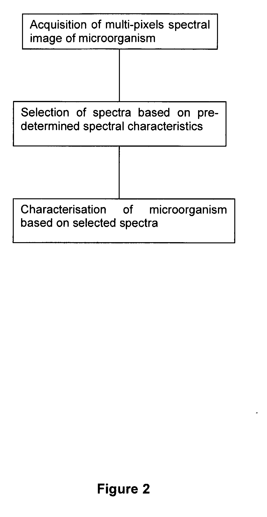
Method for the Spectral Identification of Microorganisms
a microorganism and spectral data technology, applied in the field of spectral data-based microorganism identification methods, can solve the problems of waning ir bacteria identification, time-consuming and impractical procedures, and inability to use ir spectroscopy to identify bacteria as useful techniques, etc., to achieve rapid and reliable identification.
- Summary
- Abstract
- Description
- Claims
- Application Information
AI Technical Summary
Benefits of technology
Problems solved by technology
Method used
Image

Examples
example 1
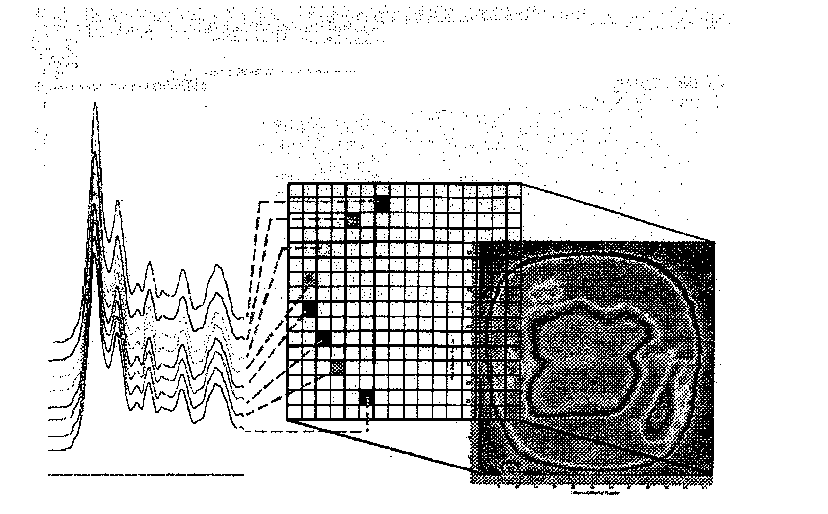
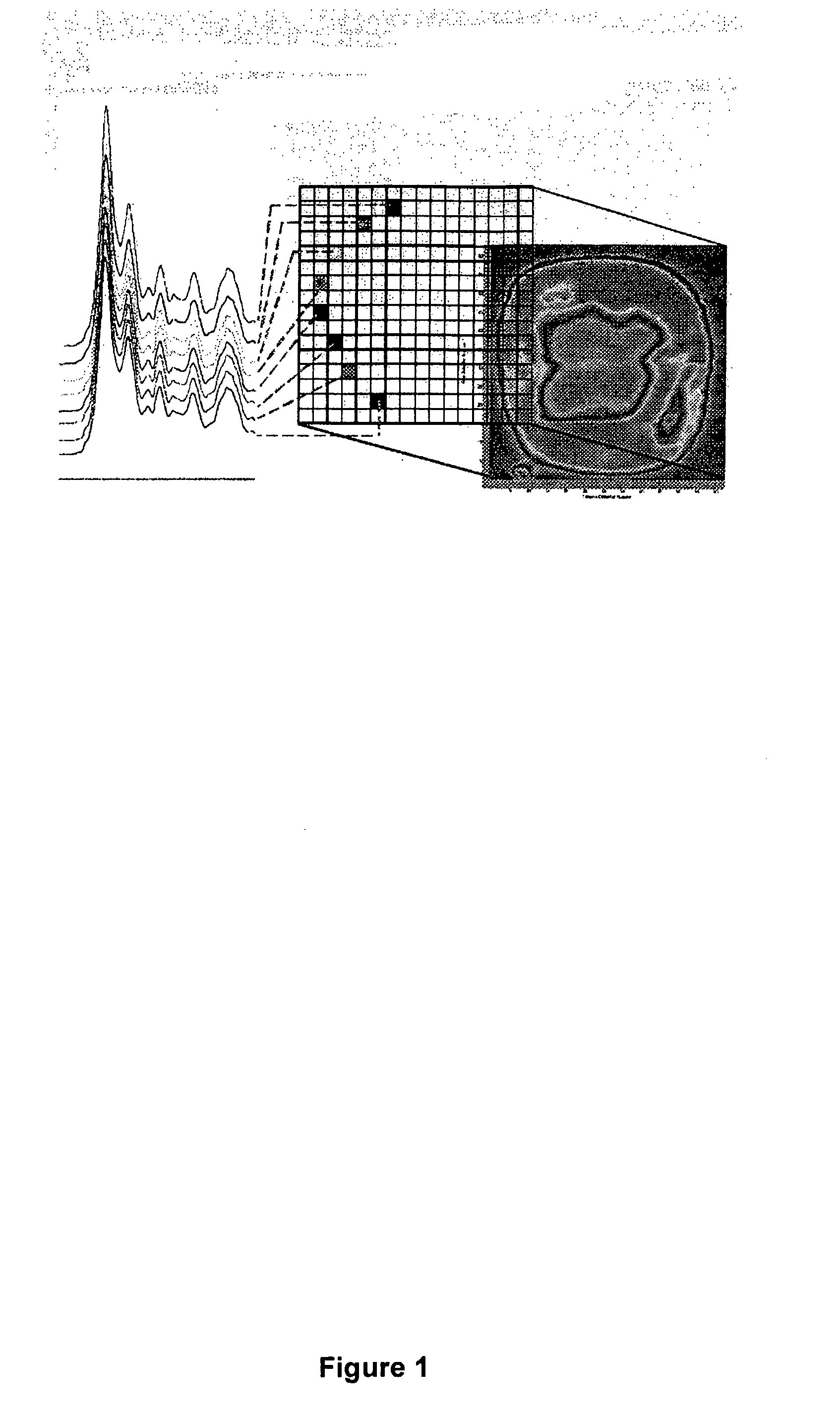
[0045]In this experiment 80 bacterial samples were characterized by FPA-FTIR. All spectra were collected on a Digilab FastIR imaging spectrometer equipped with a Digilab UMA-600 infrared microscope and a 16×16 MCT focal plane array detector (Digilab, Randolph, Mass., USA) operating under Win-IR Pro 3.3 (Digilab) by co-adding 256 scans at a resolution of 8 cm−1. In order to produce an absorbance, all single beam spectra and focal plane array imaging spectra were ratioed against the open beam spectrum of the sample. A constant flow of dry air was employed to purge the spectrometer and the microscope of carbon dioxide and water vapor. In total, (for the 80 bacterial samples) over 200,000 spectra of bacteria and bacterial cell extracts were recorded from the spectral images, yielding an average of 250 good pixels out of a possible 256 pixels per image, (each pixel generates a unique spectrum). A sterile wood toothpick was inserted into isolated colonies of bacteria to gently scrape some...
example 2
[0049]For this evaluation of FPA-FTIR spectroscopy, two Digilab Stingray systems, one equipped with a 16×16 and the other with a 32×32 array detector, were used.
Experimental Protocols
[0050]Growth of bacteria. Spectra of bacteria can be recorded from intact cells taken directly from culture plates. As early as the 1950s, it was recognized that the IR spectra of living bacterial cells strongly depend on the composition of the growth medium and time of growth. Consequently, extremely precise metabolic control and strict standardized handling of all samples is preferable to yield sufficient spectral reproducibility for comparison of the IR spectra of bacteria. To facilitate the use of FTIR spectroscopy in routine microbiological analysis, “Universal Medium” (UM™) was used. To date, this medium has supported the growth of virtually all bacteria and yeasts that have been tested. All 100 strain employed in the present work were grown on UM™ agar plates (provided by Quelab Laboratories Inc....
example 3
Materials and Methods
Microbiological Specimens
[0058]A total of 8 strains of C. botulinum (CK2 A, 2 B, 17 B, 13983 B, Bennett E, Russ E, H461297 F, and 602 F) were used in this study. This investigation was only concerned with strains of C. botulinum in groups A, B, E, and F because only these serotypes give rise to intoxications in humans. The isolates were confirmed as C. botulinum because of the production of characteristic botulinum neurotoxins.
FTIR Spectroscopic Methods
[0059]Prior to FTIR spectral acquisition, all strains were incubated for 48 hours on both Brain Heart Infusion (BHI) and McClung Toabe with egg yolk (MTEYE) media. To optimize the growth conditions, proteolytic strains were grown at 35° C., while non-proteolytic strains were grown at 25° C. The bacteria were transferred to an infrared transmitting window (ZnSe). The bacteria were air dried on the IR window for 10 minutes and then incubated in formaldehyde vapours for several hours to ensure inact...
PUM
| Property | Measurement | Unit |
|---|---|---|
| spectra | aaaaa | aaaaa |
| spectrum | aaaaa | aaaaa |
| spectral intensity | aaaaa | aaaaa |
Abstract
Description
Claims
Application Information