Medical image reporting system and method

a medical image and reporting system technology, applied in the field of medical imaging, can solve the problems of non-standard patient anatomy, exacerbate difficulties, and fail to extract small peripheral airways by fully automatic methods, and achieve the effects of low false-positive rate, strong performance, and high sensitivity to peripheral airways

Active Publication Date: 2017-06-06
PENN STATE RES FOUND
View PDF8 Cites 8 Cited by
  • Summary
  • Abstract
  • Description
  • Claims
  • Application Information

AI Technical Summary

Problems solved by technology

Fully-automated methods, however, may fail to extract small peripheral airways.
These difficulties are exacerbated when trying to define endoluminal airway-wall surfaces, especially in the airway periphery.
Additional complications arise during MDCT image reconstruction, which involves a choice of convolution kernels.
Soft kernels, such as the Siemens B3 I f kernel, have a smoothing effect and tend to blur small airways.
Motion artifacts, non-standard patient anatomy, and airway obstructions introduce additional challenges.
Choosing an appropriate global HU threshold is difficult, however, as the lungs are filled with air and misclassifying a single wall voxel can allow the segmentation to leak into the lung parenchyma.
Morphological filters are frequently slow, however, and developing an appropriate reconstruction algorithm has proven to be difficult.

Method used


Image

  • Medical image reporting system and method
  • Medical image reporting system and method
  • Medical image reporting system and method

Examples


Embodiment Construction

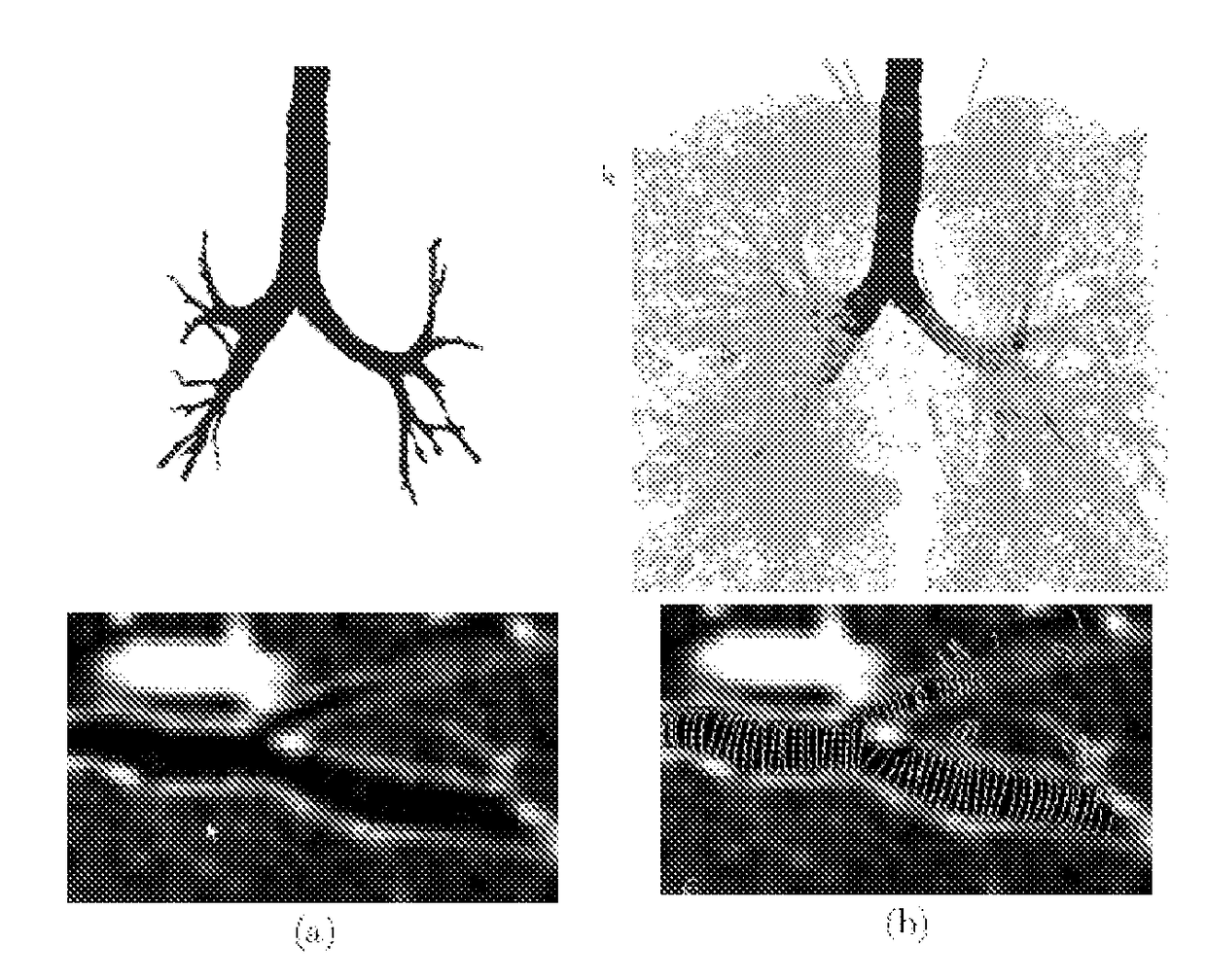
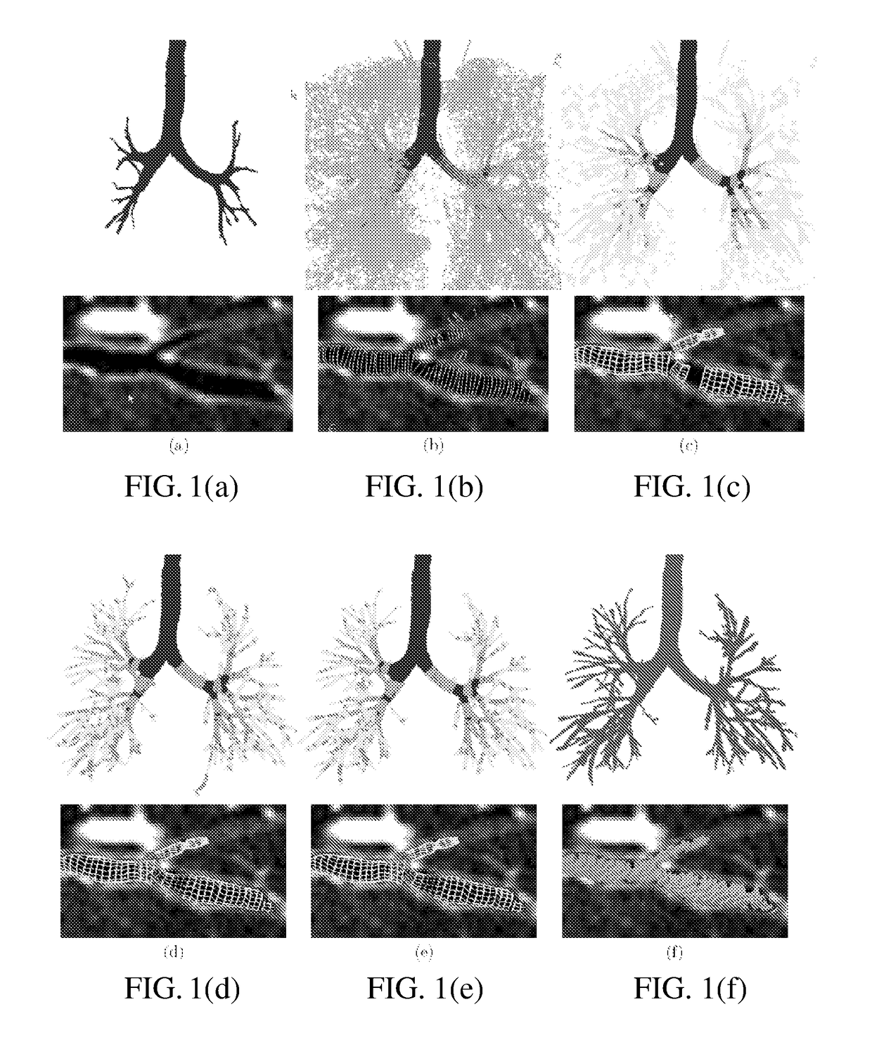
[0022]We propose a global automatic airway segmentation algorithm. The algorithm uses seeded region-growing to quickly and reliably extract the major airways. Leakage is avoided by smoothing the image. The algorithm also runs an efficient nonlinear filter over the entire lung volume in search of airway signatures. The filter output guides and constrains a locally-adaptive region-growing algorithm, which segments and connects entire airway branches. Finally, a novel graph-partitioning algorithm makes an optimal global decision on which branches to retain in the segmentation.

[0023]The proposed automatic segmentation algorithm extracts a large fraction of the visible airways with few false positive branches. For some applications, however, even this high accuracy rate can be insufficient. Consider, for example, image-based bronchoscope guidance for peripheral pulmonary lesions.29, 32 Here, VB renderings are compared to live bronchoscopic video to guide the physician along a predefined ...


PUM


Abstract

This invention relates generally to medical imaging and, in particular, to a method and system for reconstructing a model path through a branched tubular organ. Novel methodologies and systems segment and define accurate endoluminal surfaces in airway trees, including small peripheral bronchi. An automatic algorithm is described that searches the entire lung volume for airway branches and poses airway-tree segmentation as a global graph-theoretic optimization problem. A suite of interactive segmentation tools for cleaning and extending critical areas of the automatically segmented result is disclosed. A model path is reconstructed through the airway tree.

Description

REFERENCE TO RELATED APPLICATIONS[0001]This application is the United States national phase application of Patent Cooperation Treaty No. PCT / US2009 / 034197, filed Feb. 16, 2009, which claims priority from U.S. provisional patent application Ser. Nos. 61 / 028,562, filed Feb. 14, 2008; 61 / 028,607, filed Feb. 14, 2008; and 61 / 117,334, filed November 2008, the entire content of all of which are incorporated herein.STATEMENT OF GOVERNMENT SPONSORSHIP[0002]This invention was made with government support under Grant Nos. CA074325 and CA091534NIH awarded by the National Cancer Institute. The government has certain rights in the invention.BACKGROUND OF THE INVENTION[0003]This invention relates generally to medical imaging and, in particular, to a method and system for I) reconstructing a model path through a branched tubular organ, II) automatic lymph node station mapping, and III) path or route report generation. Robust and accurate reconstruction of the human airway tree from multi-detector ...

Claims


Application Information

Patent Timeline
Patent Type & Authority Patents(United States)
IPC IPC(8): G06K9/00G06T7/162G06T7/12
CPCG06T7/162G06T7/12G06T2207/20044G06T2207/30061
Inventor HIGGINS, WILLIAM E.GIBBS, JASON D.YU, KUN-CHANGGRAHAM, MICHAEL W.LU, KONGKUO
Owner PENN STATE RES FOUND
Why Eureka
Try Eureka