Method for extracting tail end bronchial tree from lung CT image

A CT image and bronchial tree technology, applied in the field of lung CT image processing, to achieve high sensitivity, reduce system complexity, and quickly identify and locate the effect

Inactive Publication Date: 2019-09-17
JILIN UNIV FIRST HOSPITAL
View PDF5 Cites 4 Cited by
  • Summary
  • Abstract
  • Description
  • Claims
  • Application Information

AI Technical Summary

Problems solved by technology

[0003] The technical problem to be solved by the present invention is to provide a method for extracting the terminal bronchial tree from lung CT images, so as to solve the defects in the prior art

Method used


Image

  • Method for extracting tail end bronchial tree from lung CT image

Examples


Embodiment Construction

[0012] The principles and features of the present invention are described below in conjunction with the accompanying drawings, and the examples given are only used to explain the present invention, and are not intended to limit the scope of the present invention.

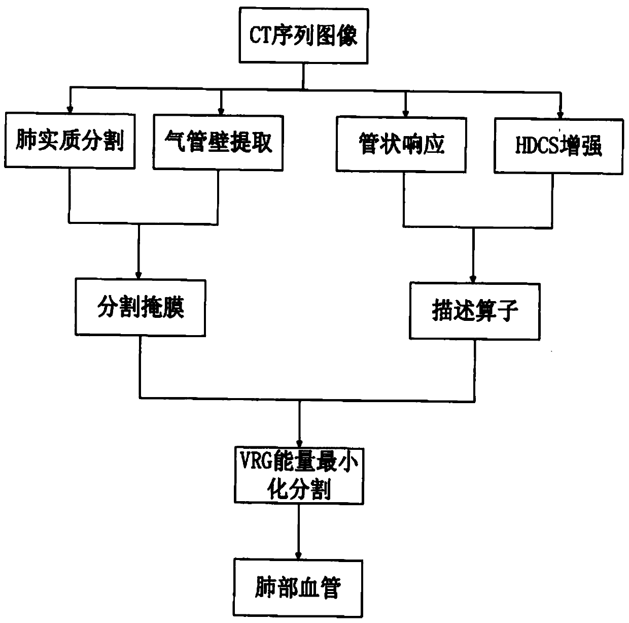
[0013] Such as figure 1 As shown, a method for extracting terminal bronchial tree from lung CT images includes the following steps:

[0014] 1) Prepare initial data: The initial data includes lung texture CT image patches used for training, verification and testing, corresponding geometric information image patches and corresponding category labels.

[0015] 2) Construction of convolutional neural network: Based on the idea of ​​skip structure in convolutional neural network, an 18-layer convolutional neural network is constructed.

[0016] 3) training based on the convolutional neural network obtained in step (2);

[0017] More specifically, the volume base layer size of the convolutional neural network is 96 con...


PUM


Abstract

The invention relates to a method for extracting a tail end bronchial tree from a lung CT image, comprising the following steps: slicing and numbering each original lung CT image according to the same sequence and size to obtain a plurality of image blocks; dividing the image blocks into diseased knots, no tissues or cavity walls, blood vessel or other lung tissues and lung cavity walls, and inputting the image blocks into a convolutional neural network for training. The image slice classification processing can reduce the system complexity, guarantees the integrity of pulmonary nodule characteristics, can be well used for the training of a convolutional neural network, has high sensitivity and low misdiagnosis rate, and enables the convolutional neural network to quickly realize the recognition and positioning of pulmonary nodules in the application.

Description

technical field [0001] The invention relates to the technical field of lung CT image processing, in particular to a method for extracting terminal bronchial trees from lung CT images. Background technique [0002] Pulmonary blood vessels are composed of pulmonary arteries and pulmonary veins, and are one of the most complex vascular structures in various tissues and organs of the human body. Starting from the pulmonary aorta and main pulmonary vein, the pulmonary blood vessels branch out step by step to form a tree-like vascular tree structure. In clinical diagnosis, accurate acquisition of the anatomical structure information of the pulmonary vascular tree is an important reference for assessing the risk of pulmonary hypertension and the basis for automatic detection of pulmonary embolism. It is also beneficial to reduce the false positive rate of pulmonary nodule detection. In clinical research, effectively separating the pulmonary vascular tree has important clinical sig...

Claims


Application Information

Patent Timeline
Patent Type & Authority Applications(China)
IPC IPC(8): G06T7/00
CPCG06T7/0012G06T2207/10081G06T2207/20081G06T2207/20084G06T2207/30061
Inventor 李丹
Owner JILIN UNIV FIRST HOSPITAL
Why Eureka
Try Eureka