Diagnostic image generation apparatus and diagnostic image generation method

a technology of diagnostic image and projection image, which is applied in the field of three-dimensional projection image generation, can solve the problems of inability to accurately extract the tissue to be imaged, difficult to distinguish between regions, and difficulty in establishing three-dimensional roi, etc., and achieves efficient interpretation of 3d image, high degree of accuracy, and increased burden on the operator

Active Publication Date: 2017-09-12
FUJIFILM HEALTHCARE CORP
View PDF5 Cites 2 Cited by
  • Summary
  • Abstract
  • Description
  • Claims
  • Application Information

AI Technical Summary

Benefits of technology

[0008]Specifically, a diagnostic image generation apparatus is provided, including a three-dimensional ROI setter configured to establish a three-dimensional ROI to which a rendering process is applied, on volume data being an aggregate of data acquired from three-dimensional space within a living body, and a projected image generator configured to execute the rendering process by using the data within the three-dimensional ROI, so as to generate a three-dimensional projected image, the three-dimensional ROI setter including, an accepting part configured to accept an instruction from a user, on a predetermined tomographic image of the volume data, and a clipping plane determiner configured to determine a clipping plane that spatially separates tissue to be imaged and tissue not imaged in the three-dimensional ROI, and the clipping plane determiner determines a boundary curve on the tomographic image, the boundary curve passing a start point that is specified by the instruction, and connecting pixels that minimize a sum of energy values obtained by reflecting a brightness value of one pixel to a brightness value of an adjacent pixel, and determines the clipping plane from the boundary curve.
[0009]In addition, a diagnostic image generation method is provided with an accepting step that accepts an instruction from a user on a predetermined tomographic image of the volume data being an aggregate of data acquired from three-dimensional space within a living body, and a clipping plane determining step that determines a clipping plane to spatially separate tissue to be imaged and tissue not imaged in the three-dimensional ROI in which the volume data is subjected to the rendering process, and a projected image generation step that executes the rendering process on the volume data in the three-dimensional ROI specified by the clipping plane, and the clipping plane determining step determines a boundary curve on the tomographic image, the boundary curve passing a start point specified by the instruction, and connecting pixels that minimize a sum of energy values obtained by reflecting a brightness value of one pixel to a brightness value of an adjacent pixel, and determines the clipping, plane from the boundary curve.
[0010]According to the present invention, in generating a three-dimensional projected image by rendering, from medical volume data, it is possible to support establishing of a desired three-dimensional ROI by a simple method with a high degree of accuracy, without increasing burden on the operator, thereby achieving more efficient interpretation of 3D image and streamlining of diagnostic flow, with the use of the diagnostic image generation apparatus.

Problems solved by technology

However, in actual volume data, it is likely to be difficult to make a distinction according to the brightness value, between a region to be imaged and a region not imaged.
Therefore, this method may fail to accurately extract the tissue to be imaged and establish the three-dimensional ROI.

Method used


Image

  • Diagnostic image generation apparatus and diagnostic image generation method
  • Diagnostic image generation apparatus and diagnostic image generation method
  • Diagnostic image generation apparatus and diagnostic image generation method

Examples


first embodiment

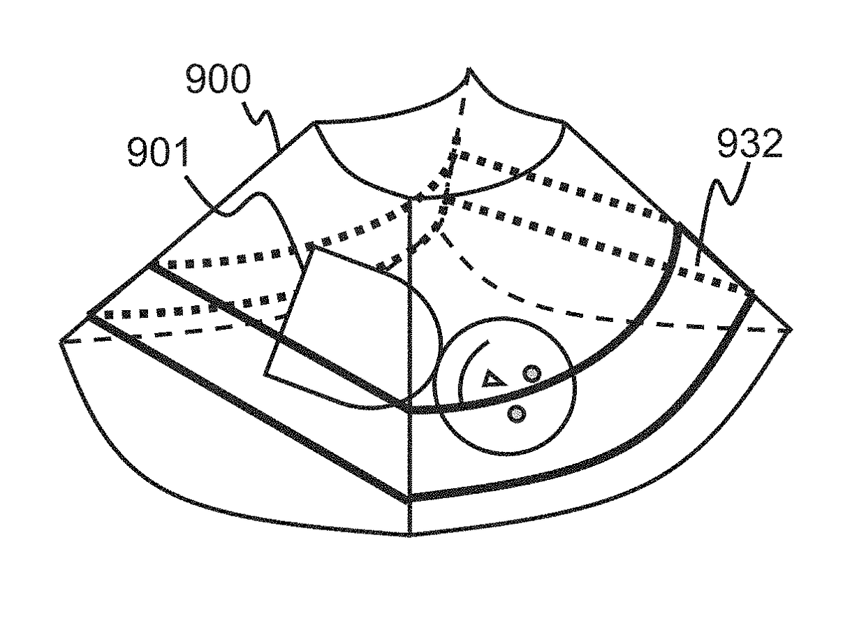
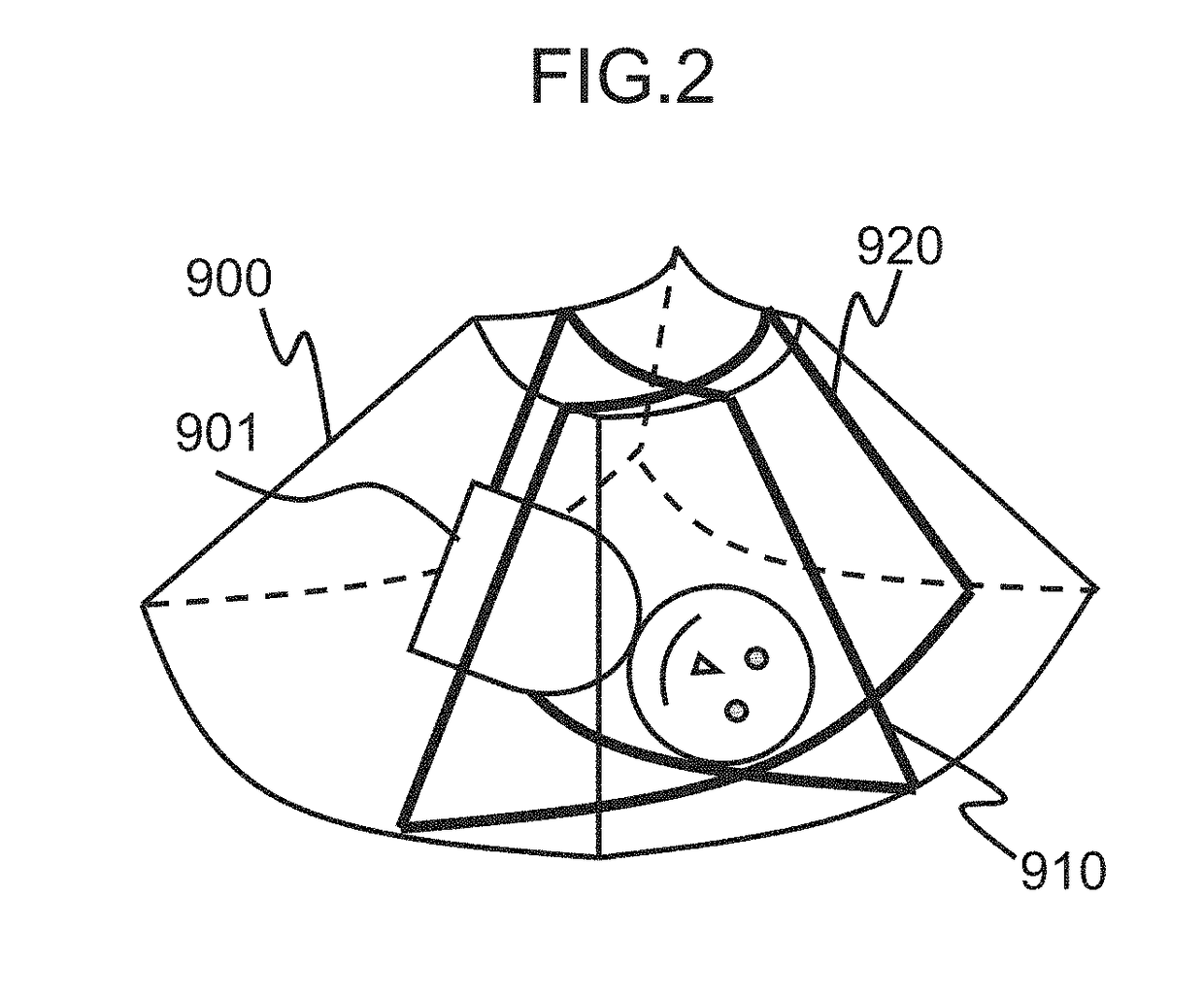
[0038]The first embodiment to which the present invention is applied will be explained. Here, an ultrasound image-capturing apparatus will be taken as an example to explain the diagnostic image generation apparatus. A fetus is assumed as the tissue to be imaged. Therefore, floating substance in amniotic fluid, placenta, and the like, may be the tissue not imaged.

[Configuration of the Ultrasound Image-Capturing Apparatus]

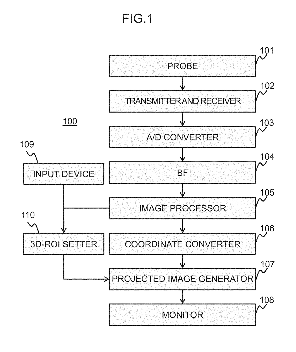
[0039]FIG. 1 is a functional block diagram showing the ultrasound image-capturing apparatus (diagnostic image generation apparatus) according to the present embodiment. As illustrated, the ultrasound image-capturing apparatus 100 of the present embodiment is provided with a probe 101 with ultrasonic transducers configured to acquire three-dimensional echo data, a transmitter and receiver 102 configured to control a transmit pulse and amplify a received echo, an analogue / digital converter (analogue / digital converting means; A / D) 103 configured to digitalize the receiv...

second embodiment

[0110]Next, a second embodiment to which the present invention is applied will be explained. In the first embodiment, the user inputs the boundary curve in the same manner as a conventional example. Then, a start point for generating the energy map is determined, on the basis of the boundary curve inputted by the user, and the boundary curve is corrected assuming the start point as the point of origin. Here, in the present embodiment, the user inputs this start point, instead of the boundary curve.

[0111]The ultrasound image-capturing apparatus 100 of the present embodiment has basically the same configuration as the first embodiment. However, since the information inputted by the user is the start point, not the boundary curve, the configuration of the three-dimensional ROI setter 110a is different. Hereinafter, the present embodiment will be explained, focusing on the configuration that is different from the first embodiment.

[0112]Also in the present embodiment, an explanation will...

modification example

[0141]As shown in FIG. 20, the three-dimensional ROI setter 110b may further be provided with a noise reducer 230 configured to perform noise reduction process.

[0142]The noise reducer 230 applies the noise reduction process to the tomographic image (here, the axial image 400) that is used in the process performed by the three-dimensional ROI setter 110b. The noise reduction process may include a smoothing process and a filtering process, such as Gaussian smoothing and Median filtering. It is desirable that a filter used in the filtering process is an edge preserving type, such as bilateral filter and Guided Filter, which removes only the noise caused by a floating substance, multiple reflection, and the like, within the amniotic fluid having a relatively low brightness value, and holds the shape information of the fetus.

[0143]The accepting part 210a presents the user the axial image 400 after the noise is reduced and then accepts the designation of the start point 411. The energy ma...


PUM


Abstract

A three-dimensional region of interest (ROI) is established with a high degree of accuracy, by a simple method without increasing a burden on the operator, in generating a three-dimensional projected image from medical volume data according to rendering, achieving more efficient interpretation of three-dimensional image and streamlining of diagnostic flow, with the use of the diagnostic image generation apparatus. An energy map is generated on a predetermined tomographic plane, assuming a preset start point as a reference and searching for a path that minimizes the energy, and then the path is set as a boundary of the three-dimensional ROI. The start point may be decided on the basis of the boundary inputted by a user, or the user may set the start point. The user may be allowed to adjust the boundary having been set. The boundary may also be determined on another plane orthogonal to the predetermined tomographic plane.

Description

INCORPORATION BY REFERENCE[0001]The present application claims priority from Japanese patent application JP-2014-0068619 filed on Mar. 28, 2014, the content of which is hereby incorporated by reference into this application.FIELD OF THE INVENTION[0002]The present invention relates to a technique for generating a three-dimensional projected image in a diagnostic image generation apparatus, and more particularly, it relates to a technique for establishing in volume data being acquired, a three-dimensional region of interest (ROI) to which a rendering process is applied.DESCRIPTION OF THE RELATED ART[0003]A diagnostic image generation apparatus, including an ultrasound diagnostic apparatus, for acquiring a medical image, is required to generate a two-dimensional image (projected image) from acquired three-dimensional image data (medical volume data), and visualize an image of tissue that is targeted for imaging. For this purpose, such apparatus is equipped with a function of generating...

Claims


Application Information

Patent Timeline
Patent Type & Authority Patents(United States)
IPC IPC(8): G06T15/08G06T3/40G06T19/00A61B8/00G06T7/181A61B8/13A61B8/08G06T7/12
CPCG06T15/08A61B8/0866A61B8/13A61B8/469A61B8/5207G06T3/40G06T7/12G06T7/181G06T19/00A61B8/466A61B8/483G06T2207/10012G06T2207/10076G06T2207/10136G06T2207/20101G06T2207/20104G06T2207/30044G06T2210/41G06T2219/008
Inventor OGINO, MASAHIRONOGUCHI, YOSHIMISHIBAHARA, TAKUMA
Owner FUJIFILM HEALTHCARE CORP
Why Eureka
Try Eureka