Flexible x-ray, detector with optical shape sensing

a detector and flexible technology, applied in the field of medical imaging, can solve the problems of not addressing the calculation of line elements of fibers or the establishment of the matrix for converting strain measurements, and achieve the effect of reducing, alleviating or eliminating on

Inactive Publication Date: 2018-06-05
KONINKLJIJKE PHILIPS NV
View PDF20 Cites 0 Cited by
  • Summary
  • Abstract
  • Description
  • Claims
  • Application Information

AI Technical Summary

Benefits of technology

[0010]It would be advantageous to achieve a system for radiation therapy where movement in the patient may be compensated for. In general, the invention preferably seeks to mitigate, alleviate or eliminate one or more of the above mentioned disadvantages singly or in any combination. In particular, it may be seen as an object of the present invention to provide a method that solves the above mentioned problems, or other problems, of the prior art.
[0011]The present principles can provide benefits such as, e.g., better feedback for a physician on location and progress of radiation therapy within an anatomy of a patient.
[0015]The optical shape sensing system is employed to track position of the target volume or target area. The present system also permits for rapid acquisition of three-dimensional (3D) volumetric sweeps of a catheter or a scope's elongated distal segment. This provides an electronic mapping or position determination of a 3D volumetric space, and provides volumetric point clouds which can facilitate registration and segmentation of intra / pre-procedurally acquired datasets.
[0019]Advantageously the sensor device comprises multiple areas of cladding distributed along a length of the sensing device. By having multiple sites or areas of cladding distributed along a part of the sensing device a better resolution is achieved.
[0022]Advantageously the optical shape sensing device includes an area of higher sensitivity by including an area with a higher number of optical fibers having optical strain sensors. E.g. optical shape sensing device may have one area where one number of optical fibers having optical strain sensors are present, in another are another number of optical fibers having optical strain sensors is present, and thus an area having of higher sensitivity may be established. The higher sensitivity may help achieve a better resolution on the determination of the position of the device. It may be advantageous to have an area having one optical fiber having optical strain sensors defining an area having a first sensitivity, and another area having four optical fibers having optical strain sensors defining an area having a second sensitivity being higher than the first sensitivity.
[0029]The method provides possibility to perform radiation therapy whilst taking into account movement in the patient, e.g. due to breathing as described above. Thereby a more efficient radiation therapy is achieved and the patient is subjected to less radiation as the supplied radiation is more efficient, this may result in less radiation therapy sessions, reduced beam width of the radiation source as the beam may be better focused during therapy and other beneficial effect of tracking movement in the target volume or area.

Problems solved by technology

However, the art fails to address how the line elements of the fiber are calculated or how the matrix for converting the strain measurements is established.

Method used


Image

  • Flexible x-ray, detector with optical shape sensing
  • Flexible x-ray, detector with optical shape sensing
  • Flexible x-ray, detector with optical shape sensing

Examples


Embodiment Construction

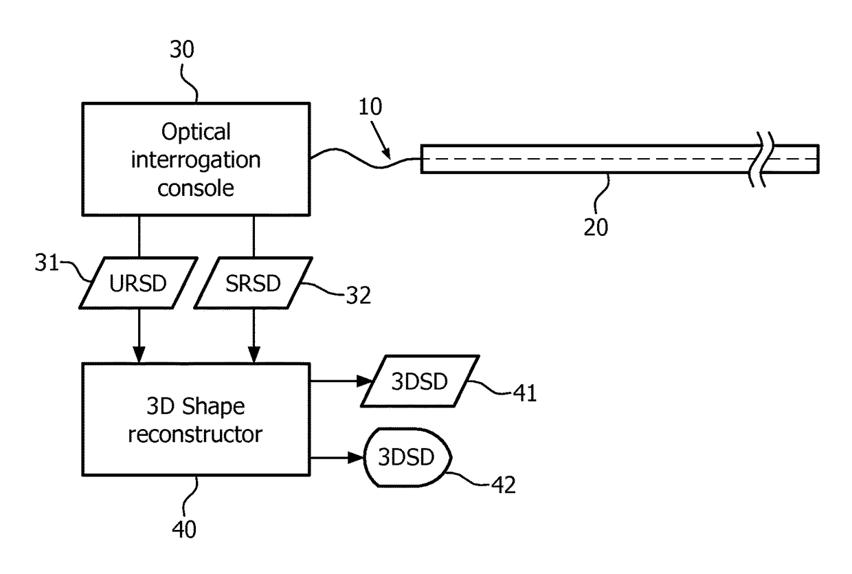
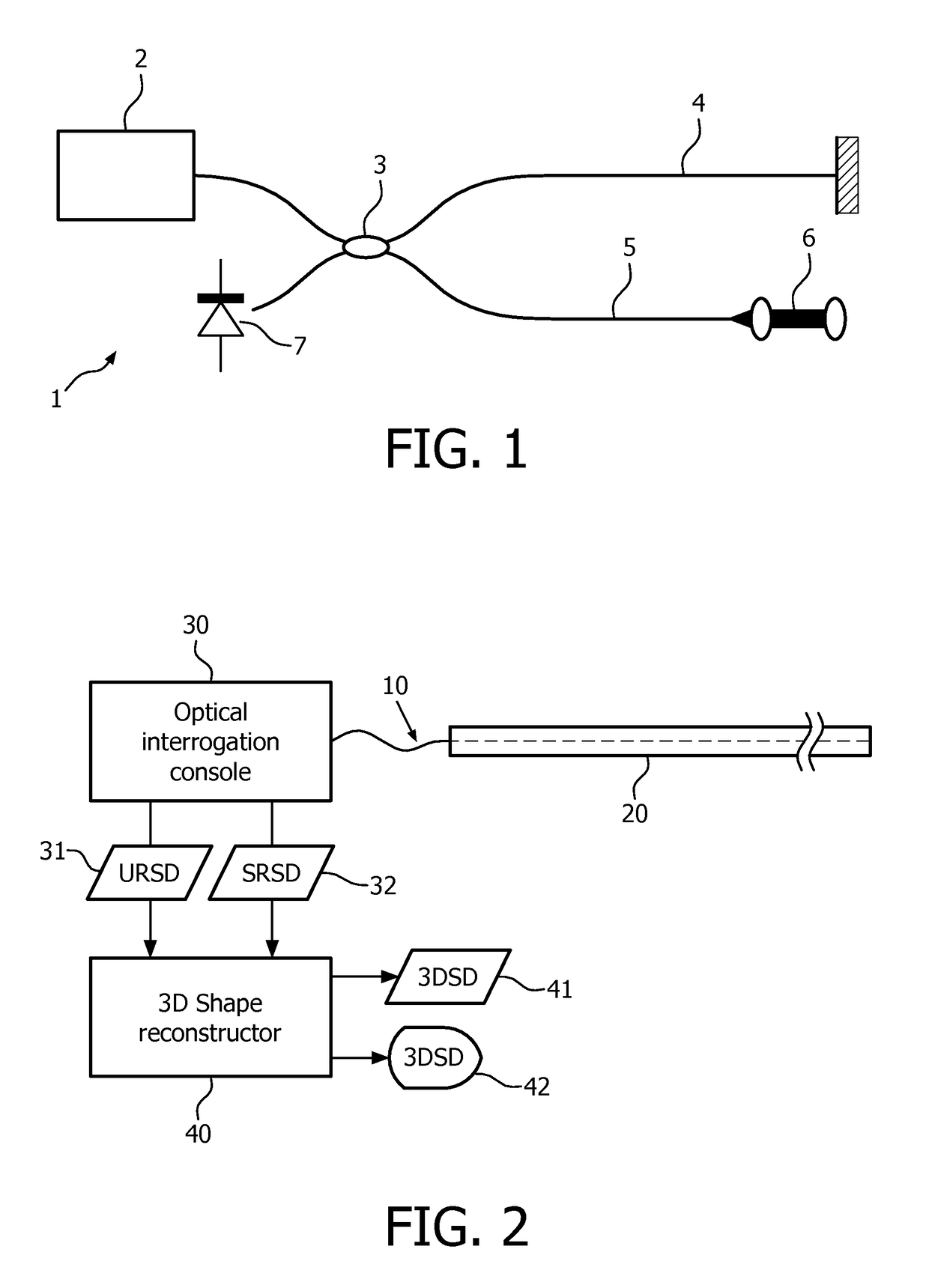
[0042]The present disclosure describes systems and methods for a radiation dosage sensing device in combination with an optical shape sensing fiber tracking system. The device may additionally designed to be imagable using a range of different ways including, but not limited to kV or MV X-ray imaging in the radiotherapy setup, or Ultrasound, further pre-interventional imaging including but not limited to CT, MR, X-ray, Ultrasound imaging could be performed. The present embodiments make use of shape reconstruction capabilities of optical sensing shape-based volumetric definition for live processing of 3D imaging data for optimising radiation treatment.

[0043]FIG. 1 schematically illustrates the principles in a configuration of a system 1 for optical frequency domain reflectometry using a tuneable light source 2 and a fiber-optic interferometer. The output of the light source 2 travels through a splitter 3 which directs a part of the signal into a reference arm 4 and the remaining part...


PUM


Abstract

The present invention relates to a sensor device for detecting dose of radiation received at the sensor device, the sensor device comprising a flexible body having a cross-section being comparatively small relative to the length of the device, a cladding at the flexible body, the cladding converting incoming radiation into visible light, and an optical shape sensing device disposed within the flexible body and configured to determine a shape of the flexible instrument relative to a reference, the shape sensing device configured to collect information based on its configuration to map an intraluminal structure during a procedure. The present invention further relates to a radiation therapy system including such a sensor device and a method of operating a radiation therapy system including such a sensor device.

Description

CROSS-REFERENCE TO PRIOR APPLICATIONS[0001]This application is the U.S. National Phase application under 35 U.S.C. § 371 of International Application Serial No. PCT / IB2012 / 055968, filed on Oct. 29, 2012, which claims the benefit of U.S. Application Ser. No. 61 / 556,315, filed on Nov. 7, 2011. These applications are hereby incorporated by reference herein.FIELD OF THE INVENTION[0002]The present invention relates to a flexible x-ray detector with optical shape sensing. This present invention relates to medical imaging, and more particularly to systems and methods for mapping internal volumes using a combination of shape sensing and images during medical procedures.BACKGROUND OF THE INVENTION[0003]In medical procedures a minimally invasive device that can be localized with a high spatial accuracy in real time in 3D and that is able to measure the number of photons reaching the tip of the device may be valuable. E.g., in 4D radiation therapy such a device may be located in the close vici...

Claims


Application Information

Patent Timeline
Patent Type & Authority Patents(United States)
IPC IPC(8): A61N5/10G01T1/161A61B6/12G01T1/02
CPCA61N5/1067A61B6/12G01T1/161G01T1/023A61N5/1071
Inventor GRASS, MICHAELMANZKE, ROBERTCHAN, RAYMOND
Owner KONINKLJIJKE PHILIPS NV
Why Eureka
Try Eureka