Ultrasonic diagnostic apparatus and the diagnostic method thereof
A diagnostic device and ultrasonic technology, applied in the directions of sonic diagnosis, infrasonic diagnosis, ultrasonic/sonic/infrasonic diagnosis, etc., can solve the problem of inability to shorten the diagnosis time, and achieve the effect of shortening the inspection time, reducing the burden, and improving the throughput
- Summary
- Abstract
- Description
- Claims
- Application Information
AI Technical Summary
Problems solved by technology
Method used
Image
Examples
Embodiment 1
[0031] First, Embodiment 1 of the present invention will be described.
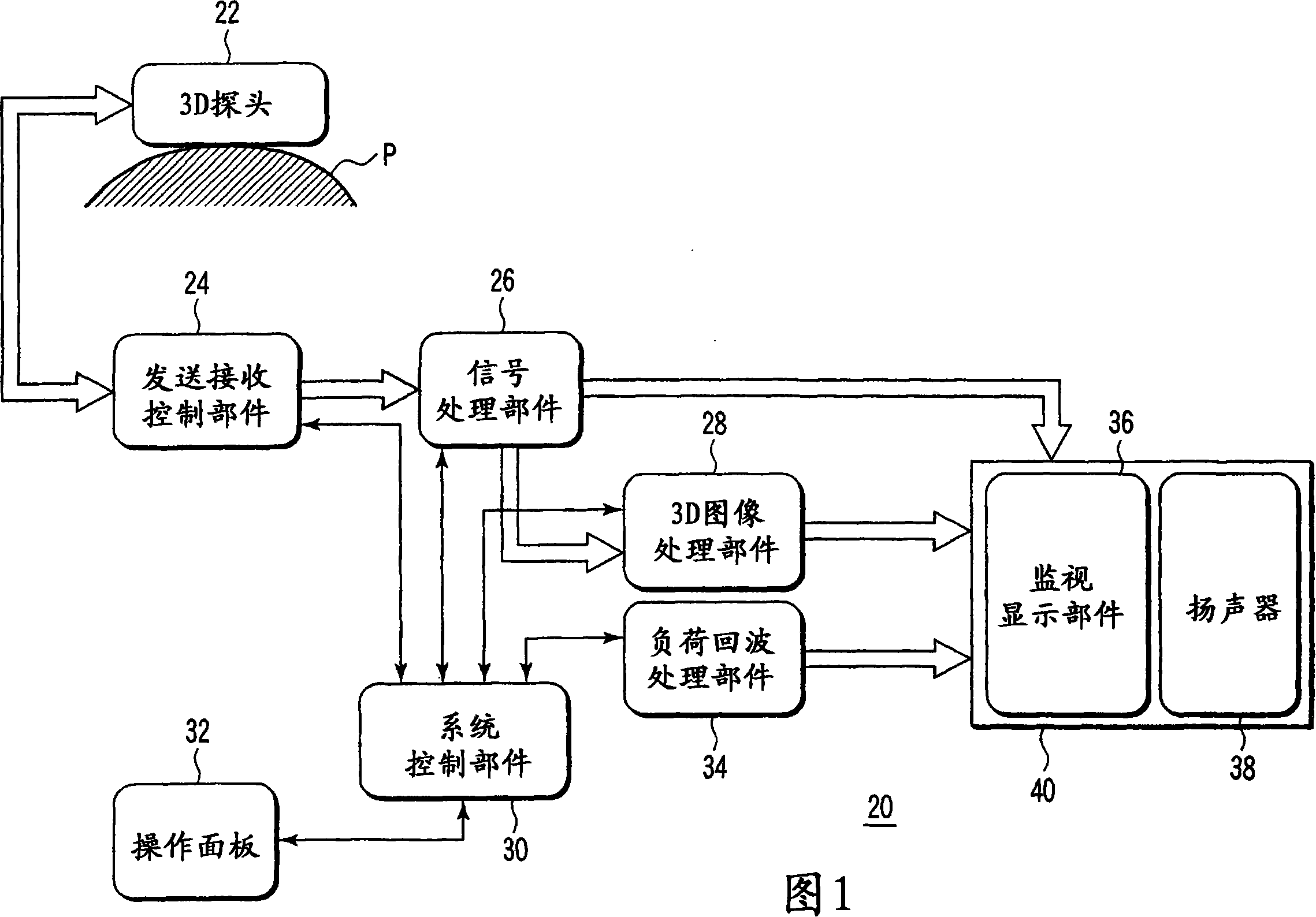
[0032] FIG. 1 is a block diagram showing a schematic configuration of an ultrasonic diagnostic apparatus according to Embodiment 1 of the present invention.
[0033] In FIG. 1 , an ultrasonic diagnostic apparatus 20 includes a three-dimensional ultrasonic probe (3D probe) 22, a transmission and reception control unit 24 including a transmission and reception unit, a signal processing unit 26, a 3D image processing unit 28, a system control unit 30, and an operation panel 32. , a load echo processing unit 34 , a monitor display unit 36 , and an output device 40 with a speaker 38 .
[0034] The 3D probe 22 transmits and receives ultrasonic waves to the subject P in order to obtain an ultrasonic tomographic image, the transmission and reception control part 24 transmits and receives electrical signals to the above-mentioned 3D probe 22, and the signal processing part 26 receives the transmission and recept...
Embodiment 2
[0065] In the first embodiment described above, the relative positional relationship between the 3D probe and the heart was shown, but in the second embodiment, a warning is issued when the tomographic image is not correctly scanned.
[0066] In addition, in this second embodiment, the structure and basic operation of the ultrasonic diagnostic apparatus are the same as those of the ultrasonic diagnostic apparatus of the first embodiment shown in FIGS. The same reference numerals are used, and illustrations and descriptions thereof are omitted, and only different parts are described.
[0067] 11 is a diagram showing an example of a monitor display layout for recognizing the positional relationship between the 3D probe 22 and the heart 50 according to the second embodiment of the present invention. Here, a warning display (for example, "warning") 100 is provided on the screen to indicate whether the four-chamber tomographic image obtained as the ROI information 74 is a correct t...
PUM
 Abstract
Description
Claims
Application Information
 - R&D
- Intellectual Property
- Life Sciences
- Materials
- Tech Scout
- Unparalleled Data Quality
- Higher Quality Content
- 60% Fewer Hallucinations
Browse by: Latest US Patents, China's latest patents, Technical Efficacy Thesaurus, Application Domain, Technology Topic, Popular Technical Reports.
© 2025 PatSnap. All rights reserved.Legal|Privacy policy|Modern Slavery Act Transparency Statement|Sitemap|About US| Contact US: help@patsnap.com


