
Ultrasonic diagnostic device

An ultrasonic diagnostic instrument and the first technology, applied in the field of ultrasonic diagnostic instruments, can solve the problems of non-adjustable transmitting circuit power, small variable gain range, and high image brightness, and achieve clear near-field images, reduce brightness, and expand The effect of gain range

Inactive Publication Date: 2008-11-19
SHENZHEN LANDWIND IND
View PDF0 Cites 14 Cited by
  • Summary
  • Abstract
  • Description
  • Claims
  • Application Information

AI Technical Summary

Problems solved by technology

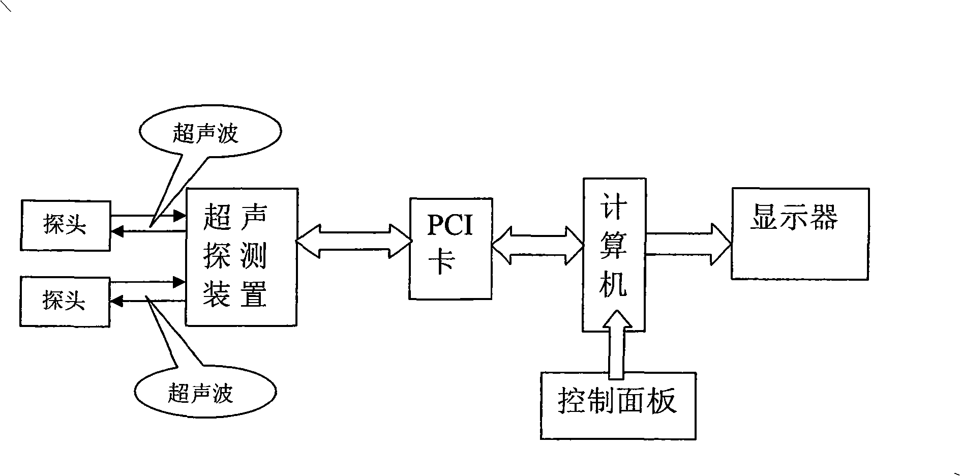
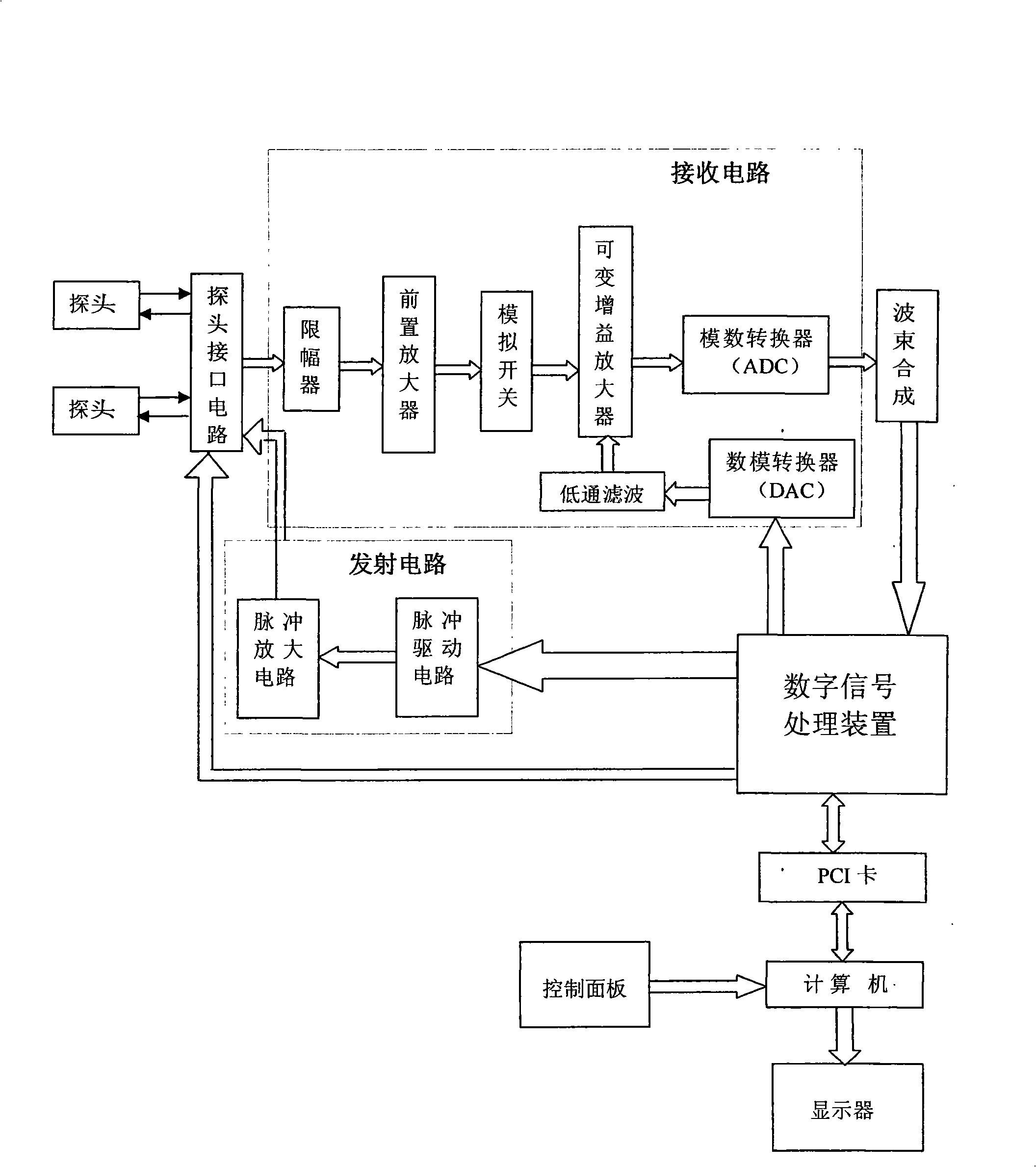
[0007] 1 There are multiple transmitting circuits corresponding to the number of probe array elements, and the power of each transmitting circuit is not adjustable, which cannot protect the fetus;
[0008] 2 The receiving module is only equipped with an adjustable gain control amplifier, and the variable gain range is small; this is very unfavorable to the echo signal processing in the near field, which will cause the image brightness in the near field to be too high, and the near field cannot be seen clearly image of

Method used


Image

  • Ultrasonic diagnostic device
  • Ultrasonic diagnostic device
  • Ultrasonic diagnostic device

Examples


Embodiment Construction

[0032] Below according to accompanying drawing and embodiment the present invention will be described in further detail:

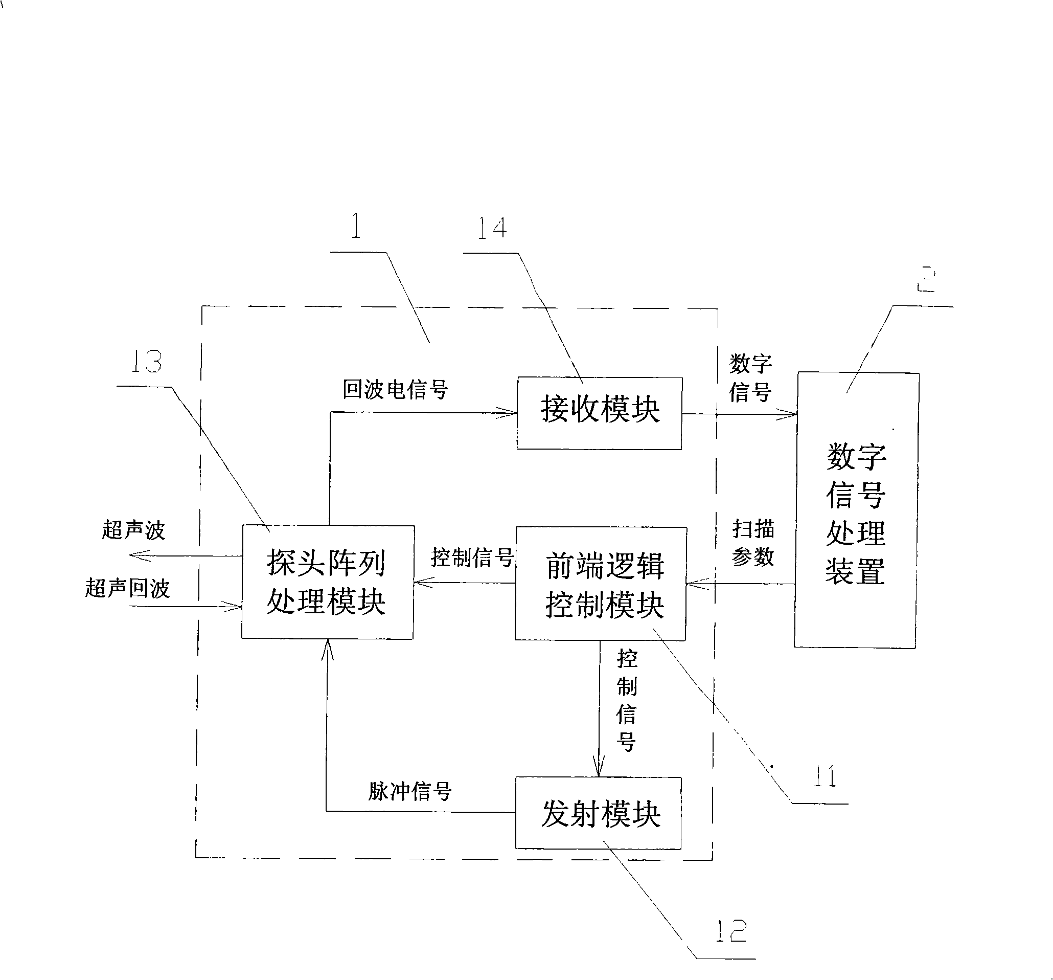
[0033] like image 3As shown, the ultrasonic diagnostic instrument of the present invention includes a front-end device 1 and a digital signal processing device 2, and the digital signal processing device 2 is used to complete phase synthesis, shift control and display superposition of digital signals; the front-end device 1 includes a front-end logic control module 11, The transmitting module 12, the probe array processing module 13 and the receiving module 14, wherein the front-end logic control module 11 is the main control module of the front-end device 1, receives the scanning parameters of the digital signal processing device 2, and completes the transmission module 12 and the probe array processing module 13 control; the transmitting module 12 sends pulse signals to the probe array processing module 13 according to the control signal of the front-en...


PUM


Abstract

The invention relates to an ultrasonic diagnostic instrument which comprises a front-end device and a digital signal processing device. The front-end device comprises a front-end logical control module, a transmission module, a probe array processing module and a receiving module; wherein, the transmission module comprises a high-pressure regulating sub-module and a transmission driving sub-module; and the receiving module comprises an amplification sub-module and an analog / digital conversion sub-module. The ultrasonic diagnostic instrument is characterized in that the amplification sub-module comprises at least a two-stage time-gain controlling amplifier. By adopting the transmission module with adjustable power, the ultrasonic diagnostic instrument is applied to various human organisms, and the damage to a fetus is avoided; a multistage time-gain controlling amplifier is arranged in the receiving module, so the gain range is expanded, and the brightness of the near field pattern is reduced, so as to lead the near field pattern to be clearer.

Description

technical field [0001] The invention relates to an ultrasonic diagnostic instrument, in particular to an ultrasonic diagnostic instrument capable of flexibly changing transmission power and multi-stage gain amplification, so as to adapt to various human tissues, avoid harm to fetuses and make near-field images clearer. Background technique [0002] Ultrasonic diagnostic equipment has become the most commonly used diagnostic equipment in medical imaging systems. Ultrasonic waves can propagate in human tissue, and the attenuation is not very large. The propagation speed of sound waves in human tissue is about 1540 meters per second. Therefore, the ultrasonic wavelength used in medical ultrasonic imaging is between 0.15 mm and 1.5 mm. Ultrasonic scanning imaging can It provides real-time images, has its own unique features in human soft tissue detection and hemodynamic observation of cardiovascular organs, and has broad development prospects. Especially in recent years, ultras...

Claims


Application Information

Patent Timeline
IPC IPC(8): A61B8/00
Inventor 林阳辉
Owner SHENZHEN LANDWIND IND
Learn More