
Molecular biology identification method of i-form duck hepatitis virus

A technology of duck hepatitis virus and molecular biology, which is applied in the field of detection, can solve problems such as technical reports on molecular biology identification methods of type I duck hepatitis virus that have not yet been seen, and achieve a wide application range, high sensitivity, and rapid diagnosis. Effect

Inactive Publication Date: 2009-02-04
SOUTH CHINA AGRI UNIV
View PDF0 Cites 12 Cited by
  • Summary
  • Abstract
  • Description
  • Claims
  • Application Information

AI Technical Summary

Problems solved by technology

[0004] There is no relevant technical report on the molecular biology identification method of type 1 duck hepatitis virus

Method used


Image

  • Molecular biology identification method of i-form duck hepatitis virus

Examples


Embodiment 1

[0035] 1. Handle samples to be tested;

[0036] (1) Tested sample

[0037] Tissue samples such as liver or kidney, cotton swabs, allantoic fluid, and cell cultures are selected, and the samples to be tested must be fresh or stored in a low-temperature refrigerator (-70°C).

[0038] A tissue sample processing

[0039] Add an appropriate amount of sterilized 0.85% normal saline to 1-3g of liver or kidney tissue samples and grind it into a 5-10 times suspension. After repeated freezing and thawing for 2-3 times, centrifuge at 4000g for 10min, and take the supernatant for nucleic acid extraction. use.

[0040] Handling of B cotton swabs

[0041] Twist the cotton swab fully and wring it dry, remove the swab, centrifuge 0.5mL sample solution at 4000g for 10min, and take the supernatant for nucleic acid extraction.

[0042] C cell culture

[0043] Take 1 mL of cell culture and freeze and thaw repeatedly 2 to 3 times, centrifuge at 4000 g for 10 min, and take the supernatant for ...


PUM


Abstract

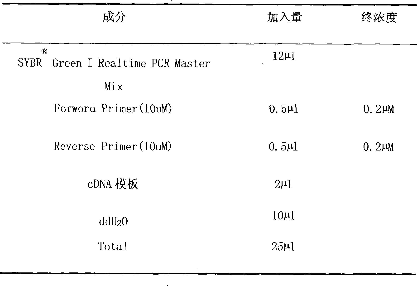
The invention discloses a molecular biological identification method for I type duck hepatitis virus, which comprises the steps of treating samples to be tested, preparing RNA of the samples, obtaining cDNA of the samples by reverse transcription, identifying I type duck hepatitis virus by the fluorescent quantitive RT-PCR method or the nested PCR method, etc. The molecular biological identification method has the advantages of fast diagnosis, high sensitivity and wide applied range, can be used for identifying DHV-1 from the tissue samples, cotton swabs, ducks or chicken embryo allantoic liquid or cell cultures.

Description

technical field [0001] The invention belongs to the technical field of detection, and in particular relates to a molecular differential diagnosis method for type I duck hepatitis virus (DHV-1). Background technique [0002] Duck viral hepatitis is a rapidly spreading and highly lethal infectious disease of ducklings. The pathological changes of the liver are characterized by increased volume and bleeding spots, and the mortality rate at the beginning of the disease is as high as more than 90%, causing great losses. [0003] Duck hepatitis is caused by 3 different viruses, duck hepatitis virus (DHV) I, II, III, the most common being DHVI, which belongs to enterovirus; DHVII is an astrovirus; DHvIII is a picornavirus . DHVI, II, and III types have obvious differences, and there is cross-immunity between each type. The size of the I virus is 20-40 nanometers, and it can grow and reproduce in the allantois of the developing chicken embryo. The virus has a strong resistance to...

Claims


Application Information

Patent Timeline
Patent Type & Authority Applications(China)
IPC IPC(8): C12Q1/70C12Q1/68C12N15/11G01N21/64
Inventor 张桂红罗玉均孔留五陈建红徐小芹廖明
Owner SOUTH CHINA AGRI UNIV
Learn More