Method for separation, characterization and/or identification of microorganisms using spectroscopy

a technology of spectroscopy and microorganisms, applied in the field of methods and systems for detecting, isolating and/or identifying microorganisms in samples, can solve the problems of bloodstream infection, high morbidity and mortality, and take several days to perform, so as to reduce the risk of handling infectious materials and/or contaminating samples, the effect of characterization and/or identification of microorganisms is faster, and the diagnosis is faster

Active Publication Date: 2010-05-27
BIOMERIEUX INC
View PDF29 Cites 56 Cited by
  • Summary
  • Abstract
  • Description
  • Claims
  • Application Information

AI Technical Summary

Benefits of technology

[0014]The present invention provides methods for isolating, characterizing and / or identifying microorganisms in a sample. The methods allow for the characterization and / or identification of microorganisms more quickly than prior techniques, resulting in faster diagnoses (e.g., in a subject having or suspected of having septicemia) and identification of contaminated materials (e.g., foodstuffs and pharmaceuticals). The steps involved in the methods of the invention, from obtaining a sample to characterization and / or identification of microorganisms, can be carried out in a very short time frame to produce clinically relevant actionable information, e.g., in less than about 120 minutes. Additionally, the methods of the invention can be fully automated, thereby reducing the risk of handling infectious materials and / or contaminating samples.
[0018]In one embodiment, the separation is carried out by layering the test sample over a density cushion in a sealed container (e.g., a hermetically sealed container) and centrifuging the container to pellet the microorganisms while the test sample medium remains on top of the density cushion. In another embodiment, the container has an optical window at the bottom and / or sides so that the microorganism pellet can be interrogated spectroscopically. The microorganisms can be identified by comparing the spectrum of the pellet to a spectrum or spectra, or spectroscopic properties predicted, of known microorganisms. The ability to identify microorganisms directly in the pellet without further handling significantly improves the safety value of this microbial identification method.

Problems solved by technology

Bloodstream infections are associated with high morbidity and mortality, yet current diagnostic methods, of culture followed by biochemical identification and antibiotic susceptibility testing, can take several days to perform.
Typically, empiric therapy is initiated based on clinical symptoms, and test results only impact clinical decisions when the initial therapy fails.
Molecular amplification methods have been proposed to fill this need, but serious challenges to this approach remain.
These direct-from-the-bottle tests are not appropriate for all microorganisms (e.g., Gram-positive cocci), are not validated by the test manufacturers, and generally take 3-8 hours to provide results.
Optical spectroscopy methods, such as intrinsic fluorescence (IF), infrared spectroscopy (FTIR), or Raman spectroscopy, and mass spectrometry methods such as MALDI-TOF, have the potential to allow for identification of microorganisms very quickly, but may encounter interference from the many highly fluorescent and absorptive compounds present in liquid microbiological culture media and in clinical samples such as blood or combinations thereof.
However, the '135 application does not discuss identification and quantification of microorganisms from complex biological samples, such as blood.
However, these methods have several drawbacks when attempting to separate and characterize microorganisms from complex samples such as blood-containing culture media.
The resultant microbial preparations often contain contaminating red blood cells, platelets, lipid particles, plasma enzymes and cellular debris, which can cause poor results.
These methods are also very labor-intensive and unsafe due to steps which can result in aerosol exposure of potentially dangerous pathogens to the user.

Method used


Image

  • Method for separation, characterization and/or identification of microorganisms using spectroscopy
  • Method for separation, characterization and/or identification of microorganisms using spectroscopy
  • Method for separation, characterization and/or identification of microorganisms using spectroscopy

Examples


example 1

Rapid Microbial Separation and Identification Method

A. Lysis-Centrifugation Separation Procedure

[0088]A suspension of colloidal silica (0.2-0.5 mL; 1.040-1.050 gm / mL density) was added to several conical microcentrifuge tubes. Lysed positive BacT / ALERT® SA blood culture broth samples (0.5-1.0 mL) were overlaid onto the colloidal silica suspension. Alternatively, the colloidal silica solution can be added underneath the lysed blood culture broth using a needle or cannula.

[0089]Positive broth from cultures containing the following microorganisms were tested:[0090]E. coli, ATCC 25922[0091]E. faecalis, ATCC 29212[0092]S. aureus, ATCC 12600[0093]P. aeruginosa, ATCC 10145

[0094]The tubes were capped, and then spun in a microcentrifuge for 2 min at about 10,000 g at room temperature (20-25° C.). The supernatants were aspirated, then the purified microbial pellets were resuspended in 0.45% w / v NaCl to an optical density @ 660 nm of 0.40.

[0095]One portion of each suspension was transferred to...

example 2

Evaluation of the Rapid Microbial Purification and Identification Method

[0098]To assess the potential of the rapid identification concept described in Example 1, twenty-four isolates (6 strains of 4 species comprising C. albicans, E. coli, S. aureus and S. epidermidis) recovered from positive blood cultures were tested in the method.

[0099]SPS-anticoagulated blood was collected from three donors and pooled. Ten mL of fresh human blood was added to BacT / ALERT® (BTA) SA blood culture bottles (bioMérieux Inc., Missouri). Suspensions of each of the isolates were prepared in tryptic soy broth (TSB). Each bottle was inoculated with 0.4 ml of a 103 / mL suspension and the bottles were incubated at 36° C. in a BTA cabinet. Four BacT / ALERT® SA bottles containing 10 mL blood but no organisms were included as negative controls.

[0100]Positive bottles were removed from the BTA cabinet within 3 hours of flagging positive. A negative control bottle was removed with each set of positive bottles (by sp...

example 3

Assessment of Lysis Buffers

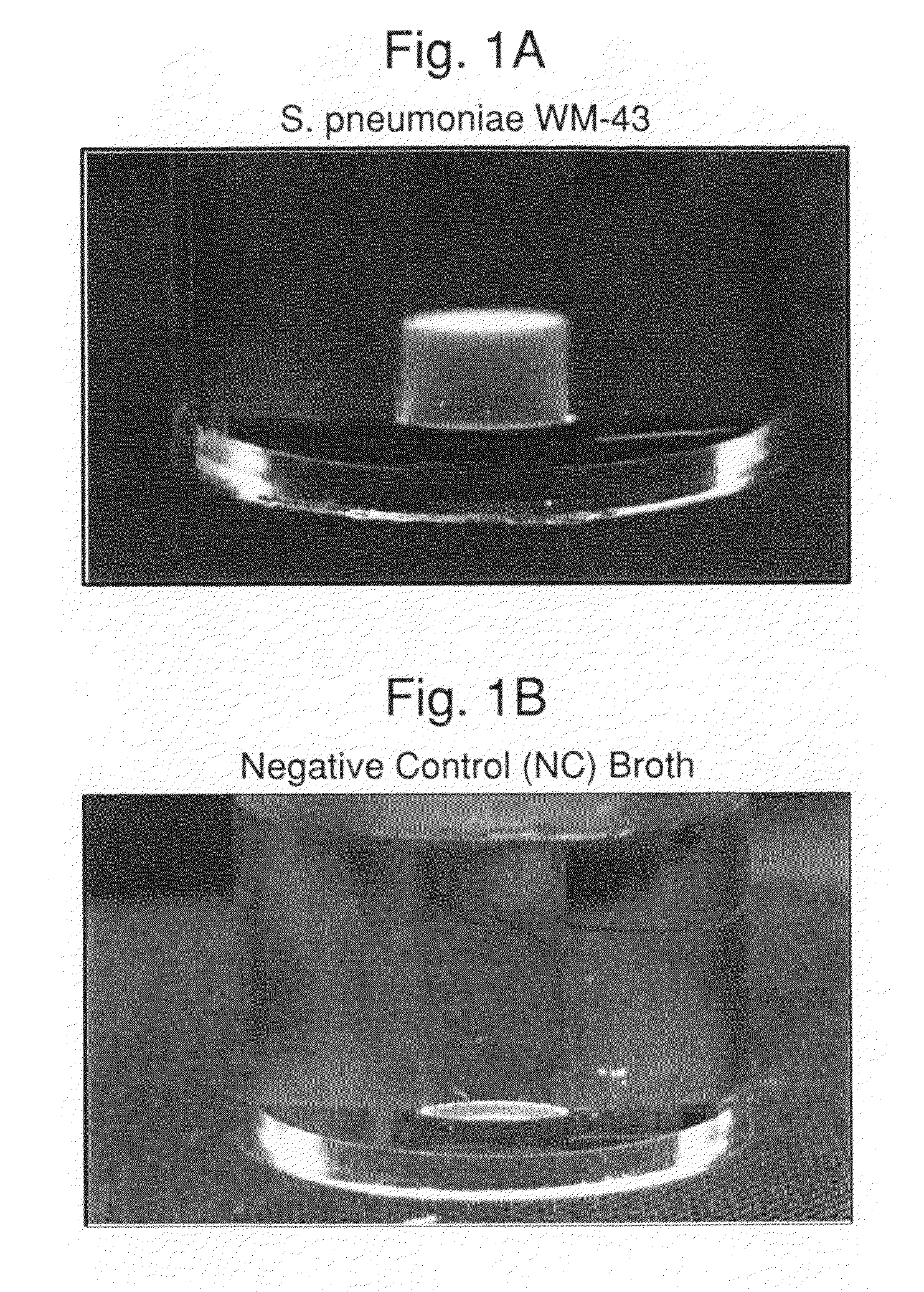
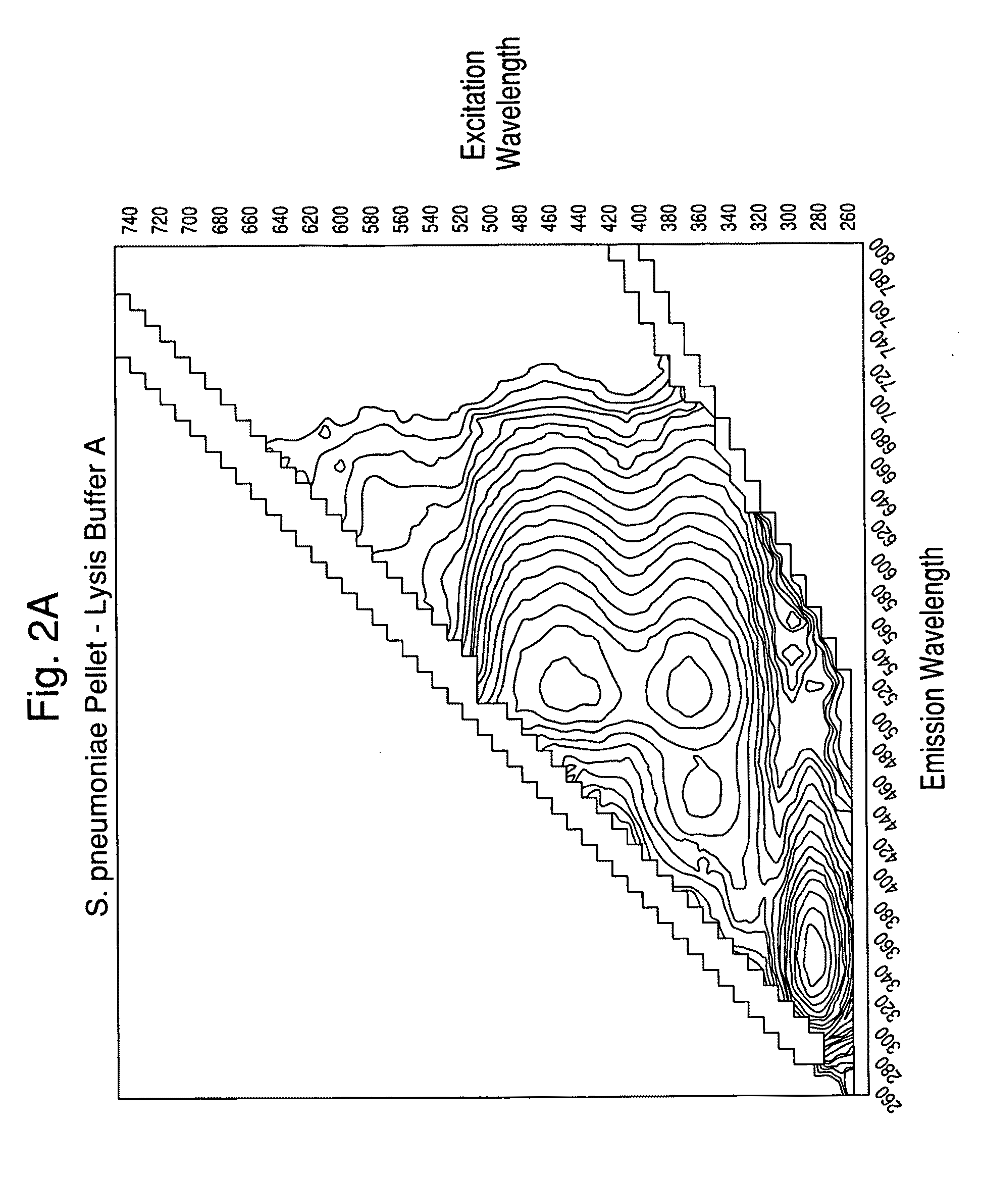
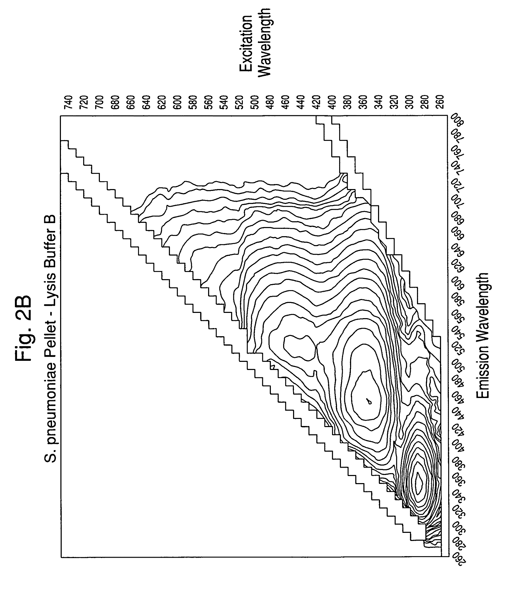
[0102]Experiments were carried out to assess the separation efficiency and microbial intrinsic fluorescence (MIF) profiles of freshly positive S. pneumoniae WM-43 blood culture broth treated with harsh and mild lysis buffers. The following lysis buffer formulations were tested: (A) 2.0% TX100-R in 0.5M CAPS, pH 11.7 (harsh=LB−A); (B) 0.75% TX100-R in 0.5M CAPS, pH 11.7 (mild=LB−B); and (C) 0.45% TX100-R in 0.3M CAPS, pH 11.7 (mild=LB−C). 1.0 mL of lysis buffers A and B and 2.0 mL of lysis buffer C were placed in screw-capped tubes and the tubes were placed in a rack in a 37° C. water bath. Samples of broth (4.0 mL) were removed from a S. pneumoniae WM43-positive BacT / ALERT® SA culture bottle using a 5 mL syringe and 18G needle within 5 minutes of flagging positive in the BTA system. The broth was quickly dispensed to each lysis buffer-containing tube and the tubes were capped and vortexed for approximately 5 seconds. A test broth from overfilled (15 mL blo...


PUM

PropertyMeasurementUnit
temperatureaaaaaaaaaa

Abstract

The present invention is directed to a method for separating, characterizing and / or identifying microorganisms in a test sample. The method of the invention comprises an optional lysis step for lysing non-microorganism cells that may be present in a test sample, followed by a subsequent separation step. The method may be useful for the separation, characterization and / or identification of microorganisms from complex samples such as blood-containing culture media. The invention further provides for spectroscopic interrogation of the separated microorganism sample to produce measurements of the microorganism and characterizing and / or identifying the microorganism in the sample using said spectroscopic measurements.

Description

CROSS REFERENCE TO RELATED APPLICATION[0001]This application claims the benefit of U.S. Provisional Patent Application No. 61 / 110,187, entitled, “Method and System for Detection and / or Characterization of a Biological Particle in a Sample”, filed Oct. 31, 2008, which is incorporated herein.FIELD OF THE INVENTION[0002]The present invention relates to methods and systems for detecting, isolating and / or identifying microorganisms in a sample. In particular, the present invention is directed to a method for rapid characterization and / or identification of a microorganism using spectroscopic techniques.BACKGROUND OF THE INVENTION[0003]The detection of pathogenic microorganisms in biological fluids should be performed in the shortest possible time, in particular in the case of septicemia for which the mortality remains high in spite of the broad range of antibiotics which are available to doctors. The presence of biologically active agents such as a microorganism in a patient's body fluid,...

Claims


Application Information

Patent Timeline
Patent Type & Authority Applications(United States)
IPC IPC(8): C12Q1/04
CPCC12Q1/04G01N21/35G01N21/3581G01N21/359G01N21/64G01N21/65G01N33/6848G01N21/47Y02A50/30C12M1/34C12M1/12
Inventor WALSH, JOHNHYMAN, JONES M.THORPE, THURMANCLAY, BRADFORDRONSICK, CHRISTOPHER
Owner BIOMERIEUX INC
Why Eureka
Try Eureka