
Method for a device for anastomosis

A technology of anastomosis and instruments, which is applied in the field of instruments for pressurized anastomosis, and can solve problems such as fistula and difficulties

Active Publication Date: 2009-05-06
CARPONOVUM AB
View PDF1 Cites 9 Cited by
  • Summary
  • Abstract
  • Description
  • Claims
  • Application Information

AI Technical Summary

Problems solved by technology

Since the two pieces of the tubular organ are placed on top of each other between the two parts of the assembly, it is difficult to evenly place the tubular organ into the assembly during installation, with the risk of fistula

Method used


Image

  • Method for a device for anastomosis
  • Method for a device for anastomosis
  • Method for a device for anastomosis

Examples


Embodiment Construction

[0049] Dehiscence of intestinal anastomosis is associated with high morbidity and mortality. Rapid and effective healing of intestinal anastomosis wounds is critical to the safety and speedy recovery of patients undergoing anastomoses. Wound healing is a relatively fixed tissue response with a cascade of predictable outcomes, including acute inflammation, hyperplasia (cell division and matrix protein synthesis), and several reorganizations of the tissue to adapt the new tissue to mechanical requirements.

[0050] A unique feature of the anastomotic device is that the wound healing process begins with local tissue ischemia and necrosis that results in the loss of previously unprocessed tissue (i.e., each separate segment of the tubular structure joined by the anastomotic ring). Segmental enteric chorion) are held together by the healing response produced by the configuration of the anastomotic instrument. It should be noted that in the abdominal cavity of a healthy person, the...


PUM


Abstract

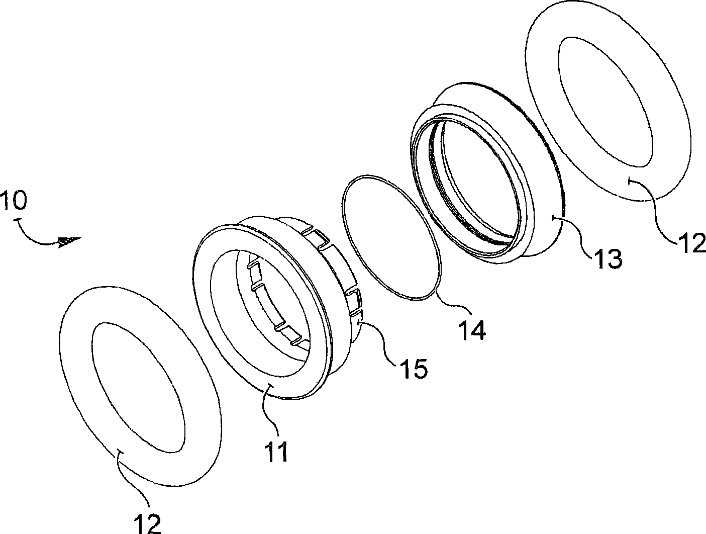
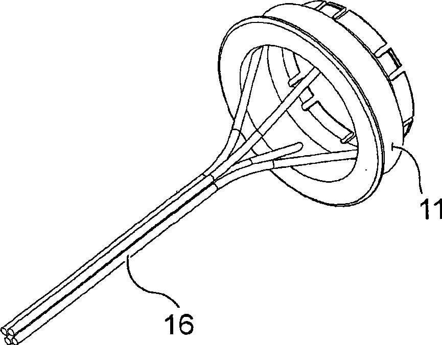
A device (10, 200) for anastomosis of a tubular structure (90) comprises a first and a second member of a generally hollow open configuration, which each includes a rigid part (11, 13) and an elastic part (12), and a connection member (15, 160) for interlocking the first and second members. According to a first embodiment the connection member (15) is integral with the rigid part (11) of the first member, and has longitudinal slits (37) forming tongues (38) there between, of which at least one has an outward protrusion (39) at its free end. ). In an alternate embodiment of the first embodiment, the connection member (15) has at least one circumferential groove (192) around the outer periphery, and the second rigid part (193) has a protrusion (194) arranged around the inner periphery. According to a second embodiment (200) the connection member (160) is arranged as a member separated from the first rigid part (11). The elastic parts (12) are essentially circular rings, and are made of a polymeric, a biocompatible and / or biodegradable material. The rigid parts (11, 13) have an outer surface that is partly semi-circular in cross section, wherein the diameter at a non-connecting end (30, 40) is larger than or equal to the diameter at a connecting end (31, 41), which ends in an edge (36, 46). The first and second members are connected to each other so that a distance is formed between the elastic parts (12). A cavity (203) is formed between the rigid parts (13) and the connection member and the tubular structure (90), when arranged in the device (200).

Description

technical field [0001] The present invention relates to an anastomosis of living tissue, and more particularly, to a device for pressurized anastomosis of tubular structures. Furthermore, the invention also relates to a method for mounting the instrument to a tubular structure. Background technique [0002] Rectal cancer is the third most frequently occurring cancer type in the world, with approximately 1 million new cases occurring each year. Carcinogenesis occurs frequently in industrial regions of the world. [0003] Current techniques for mechanically performing hollow organ anastomoses employ circular mechanical staplers [0004] (stapler), which uses metal or plastic staples (staples) to join the tissue edges of open hollow organs. Various surgical staplers have been developed for gastric surgery, esophageal surgery, and intestinal surgery. In surgical anastomosis, two parts of a hollow organ are joined, usually by a closed circular stapler, with a ring of staples....

Claims


Application Information

Patent Timeline
Patent Type & Authority Applications(China)
IPC IPC(8): A61B17/11
CPCA61B17/11A61B17/1114A61B2017/1132A61B2017/1117A61B2017/00004A61B17/12
Inventor A·格伦贝格H·索尔拉休斯
Owner CARPONOVUM AB
Learn More