Extraction method of main blood vessels from abdominal CT images based on watershed of three-dimensional region

A CT image, three-dimensional area technology, applied in the field of medical image processing and clinical applications, can solve the problems of large time complexity, reduce computational complexity, long time, etc., and achieve the effect of improving reliability and avoiding time complexity.

Inactive Publication Date: 2009-10-07
XIDIAN UNIV
View PDF0 Cites 16 Cited by
  • Summary
  • Abstract
  • Description
  • Claims
  • Application Information

AI Technical Summary

Problems solved by technology

This method can get a better vascular topology, but its time complexity is large, and it takes a long time to run
Most of the three-dimensional segmentation methods have the problems of complex models and too much segmentation time complexity, so there are quite a few blood vessel segmentation algorithms that only extract and analyze a certain part or a certain segment of the blood vessel of interest, thereby reducing the computational comple

Method used


Image

  • Extraction method of main blood vessels from abdominal CT images based on watershed of three-dimensional region
  • Extraction method of main blood vessels from abdominal CT images based on watershed of three-dimensional region
  • Extraction method of main blood vessels from abdominal CT images based on watershed of three-dimensional region

Examples


Embodiment Construction

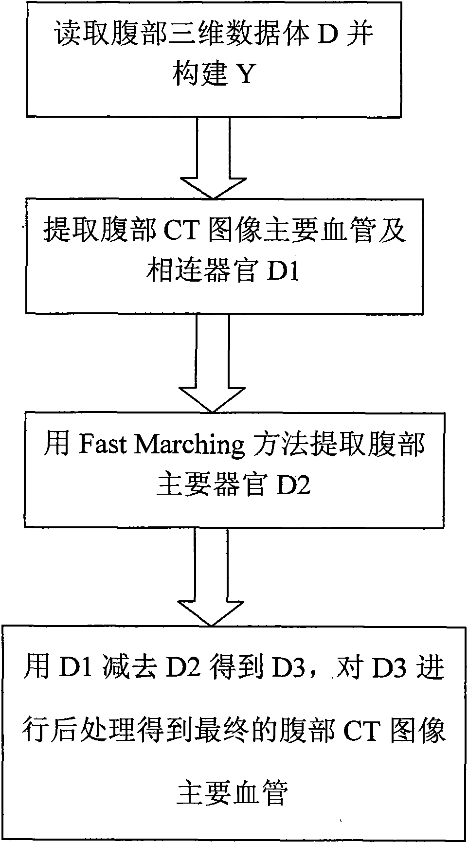
[0025] refer to figure 1 , the specific implementation steps of the present invention are as follows:

[0026] Step 1: Obtain the abdominal three-dimensional data volume D from a set of abdominal CT images;

[0027] Input a set of abdominal CT images from Beijing Cancer Hospital, 64-slice spiral CT (GE Lightspeed 64), arrange these images according to the imaging sequence, obtain the abdominal three-dimensional data volume D, and generate a new three-dimensional data with the same size as D Body Y.

[0028] The imaging sequence is a parameter recorded by the device during the contrast imaging process, and can be obtained directly from the parameter list of the CT image.

[0029] Step 2: Extract the main blood vessels and connected organs D1 from the abdominal CT image;

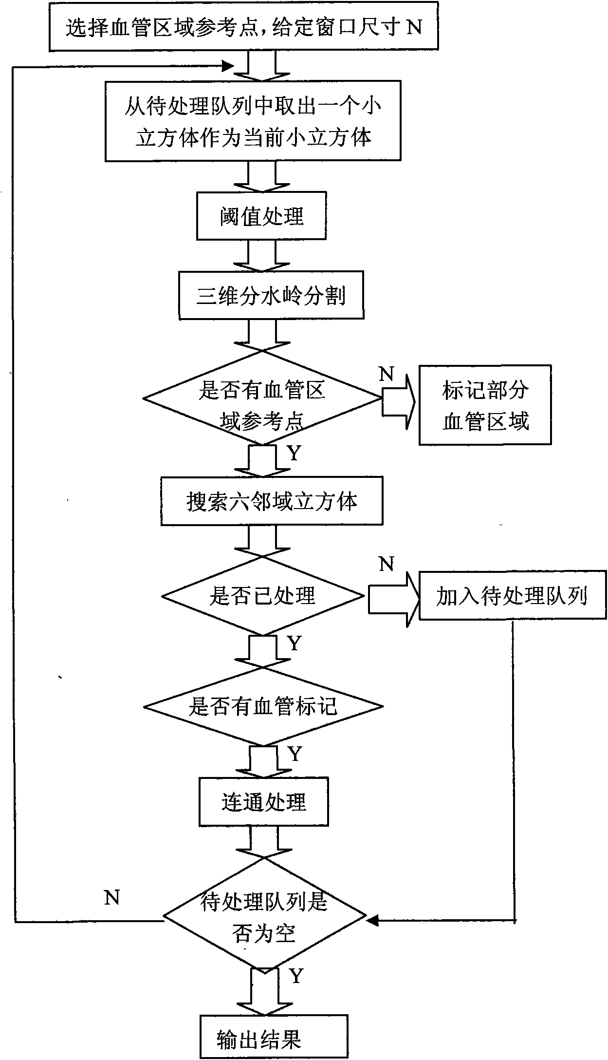
[0030] refer to figure 2 , the step is implemented as follows:

[0031] 2a. Designate a point in the abdominal three-dimensional data volume D as the reference point of the blood vessel area. The selecti...


PUM


Abstract

The invention discloses an extraction method of main blood vessels from abdominal CT images based on watershed of three-dimensional region, which mainly overcomes the shortages that the existing segmentation method of blood vessels has high complexity for computation and can not utilize the third dimension information among CT images well. The extraction method comprises the following steps of: (1) reading a set of abdominal CT images, arranging the abdominal CT images according to the imaging sequence to obtain an abdominal three-dimensional data volume; (2) implementing three-dimensional watershed cutting on a plurality of small cubes in the three-dimensional data volume, and communicating each cut small cube to extract main blood vessels and the communicated organs in the abdominal CT images from the three-dimensional data volume; (3) extracting the main abdominal organs from the three-dimensional data volume; and (4) subtracting the results respectively obtained from the step (2) and step (3), and implementing post processing on the subtracted result to obtain the final main blood vessels in the abdominal CT images. The method can faster and more completely obtain the main blood vessels in the abdominal CT images and can be used for auxiliary diagnosis of the main abdominal blood vessels.

Description

technical field [0001] The invention belongs to the field of medical image processing and clinical application, and relates to a method for extracting main blood vessels from abdominal CT images based on three-dimensional area watershed. This method can be used in abdominal CT images to extract main blood vessels to assist medical diagnosis. Background technique [0002] Segmentation of blood vessels plays an extremely important role in many practical applications of vascular diseases, such as diagnosis of stenosis or malformation, surgical planning, and surgical guidance. At present, the diagnosis of vascular diseases is mainly through angiography technology, using angiography equipment to obtain vascular images, and then doctors directly read the vascular images taken by the angiography equipment, and qualitatively judge the patient's condition based on experience and observation. At present, the post-processing of medical images using computer technology, computer graphi...

Claims


Application Information

Patent Timeline
IPC IPC(8): G06T7/00G06T15/00A61B6/03
Inventor 周伟达张莉武彩丽邹海双焦李成
Owner XIDIAN UNIV
Why Eureka
Try Eureka