Ultrasonic frequency spectrum offset parameter imaging method used for characterization of spongy bone microstructure

A spectral shift, cancellous bone technology, applied in the field of ultrasound medicine, which can solve problems such as excessive dependence on measurement position

Inactive Publication Date: 2010-03-03
FUDAN UNIV
View PDF0 Cites 14 Cited by
  • Summary
  • Abstract
  • Description
  • Claims
  • Application Information

AI Technical Summary

Problems solved by technology

(2) The transmission method uses dual probes for measurement and is too dependent on the measurement position
However,

Method used


Image

  • Ultrasonic frequency spectrum offset parameter imaging method used for characterization of spongy bone microstructure
  • Ultrasonic frequency spectrum offset parameter imaging method used for characterization of spongy bone microstructure
  • Ultrasonic frequency spectrum offset parameter imaging method used for characterization of spongy bone microstructure

Examples


Embodiment

[0030] The following takes an isolated bovine tibia sample as an example to introduce the entire process of the ultrasonic backscatter imaging method, and finally compare the backscatter image with the μ-CT image to show the results.

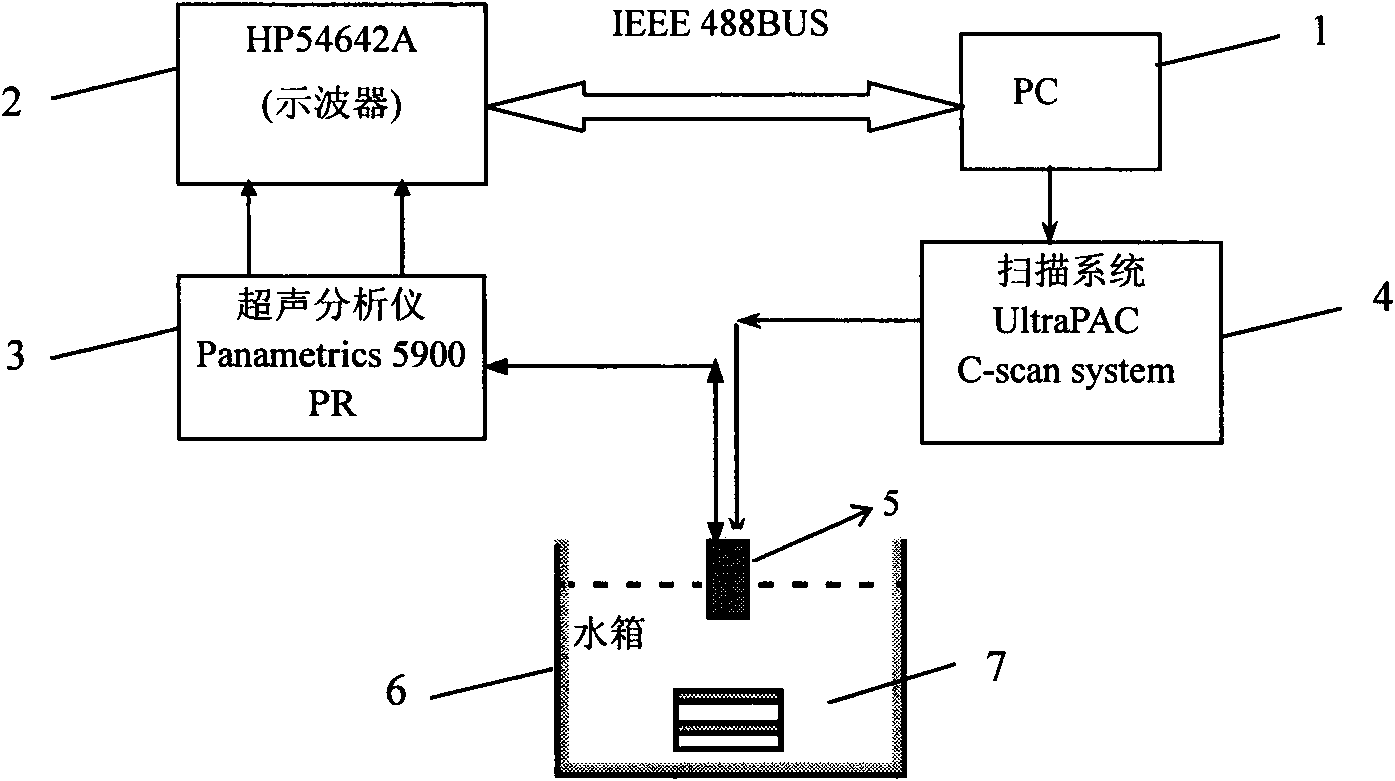
[0031] 1. Hardware design and parameter estimation

[0032] An example is given to illustrate the hardware design and parameter estimation method of ultrasonic frequency shift parameter imaging. The block diagram of the hardware system is as figure 1 shown. In the system, the ultrasonic probe is fixed at the bottom of a metal catheter, and the metal catheter can be scanned in a grid at equal intervals on the x-y plane under the automatic control of the scanning system (UltraPAC C-scansystem, USA), with a scanning step size of 0.14 mm. The parameters of the focused ultrasound probe (Panametrics, Walthan, MA) used were: center frequency 10 MHz, focal length 38.1 mm, focal column length 0.9 mm, focal diameter 0.54 mm, bandwidth 6.58-14.16 MHz. A...


PUM


Abstract

The invention belongs to the ultrasonic mechanical technical field, and particularly relates to an ultrasonic frequency spectrum offset parameter imaging method used for characterization of a spongy bone microstructure. The method comprises the following steps: scanning a spongy bone to be detected with a single high-frequency ultrasonic probe, and vertically emitting an ultrasonic pulse wave to the spongy bone at each scanning position; receiving an ultrasonic backscattering signal by the same probe when ultrasonic wave transmission is affected by the spongy bone microstructure; respectivelyperforming signal processing on the received signal at each scanning position, and extracting the frequency spectrum offset parameters of the received signal and the transmit signal to obtain an ultrasonic frequency spectrum offset parameter matrix corresponding to a scanning area; and mapping the parameter matrix into an ultrasonic frequency spectrum offset parameter image, and proportionally setting pixel values and parameter values. The method for ultrasonic imaging on the spongy bone with the frequency spectrum offset parameters of the backscattering signal is firstly proposed in the invention, and the method can help better reflect spongy bone microstructure characteristics compared with the traditional spongy bone ultrasonic imaging method based on a transmission method.

Description

technical field [0001] The invention belongs to the technical field of ultrasonic medicine, and in particular relates to an ultrasonic imaging method for evaluating the state of cancellous bone. Background technique [0002] Osteoporosis, a skeletal disease caused by decreased bone strength, is a major health concern today. The diagnosis of osteoporosis has been paid more and more attention by the academic and medical circles at home and abroad, and the imaging method of cancellous bone tissue can be used to characterize the bone condition. Compared with X-ray-based cancellous bone imaging methods, ultrasound technology has the advantages of low cost, no ionizing radiation, and simplicity. Ultrasonic parametric imaging technology of cancellous bone has attracted extensive attention. [0003] Ultrasound imaging technology that has been proposed so far is ultrasound parametric imaging based on ultrasound transmission, which scans the cancellous bone of the calcaneal bone and ...

Claims


Application Information

Patent Timeline
IPC IPC(8): A61B8/08
Inventor 他得安黄凯王威琪杨白丁
Owner FUDAN UNIV
Why Eureka
Try Eureka