Neural network-based method for identifying and classifying visible components in urine

A neural network, recognition and classification technology, applied in the field of image processing, can solve the problems of low false positive rate and identification of formed components, and achieve the effect of overcoming instability, improving accuracy and low false positive rate.

Active Publication Date: 2010-05-26
DIRUI MEDICAL TECH CO LTD
View PDF0 Cites 45 Cited by
  • Summary
  • Abstract
  • Description
  • Claims
  • Application Information

AI Technical Summary

Problems solved by technology

[0004] The present invention provides a method for identifying and classifying formed components in urine based on a neural network to solve the problem that it is difficult to identify various formed components in urine sample images with high precision and low false positive rate in the prior art

Method used


Image

  • Neural network-based method for identifying and classifying visible components in urine
  • Neural network-based method for identifying and classifying visible components in urine
  • Neural network-based method for identifying and classifying visible components in urine

Examples


Embodiment Construction

[0042] Glossary:

[0043] Urinary sediment: Refers to the formed components in urine, such as red blood cells, white blood cells and bacteria in urine.

[0044] Formed components of urine: Refers to substances such as red blood cells, white blood cells, and bacteria in the urine.

[0045] Urine sediment testing equipment: It is a clinical testing equipment for detecting formed components in urine.

[0046] Laminar flow: Laminar flow refers to the orderly flow of fluid microgroups without mixing with each other.

[0047] Flow cell: It is composed of a specially made thin-layer plate, and the detection sample forms a laminar flow under the action of the sheath fluid.

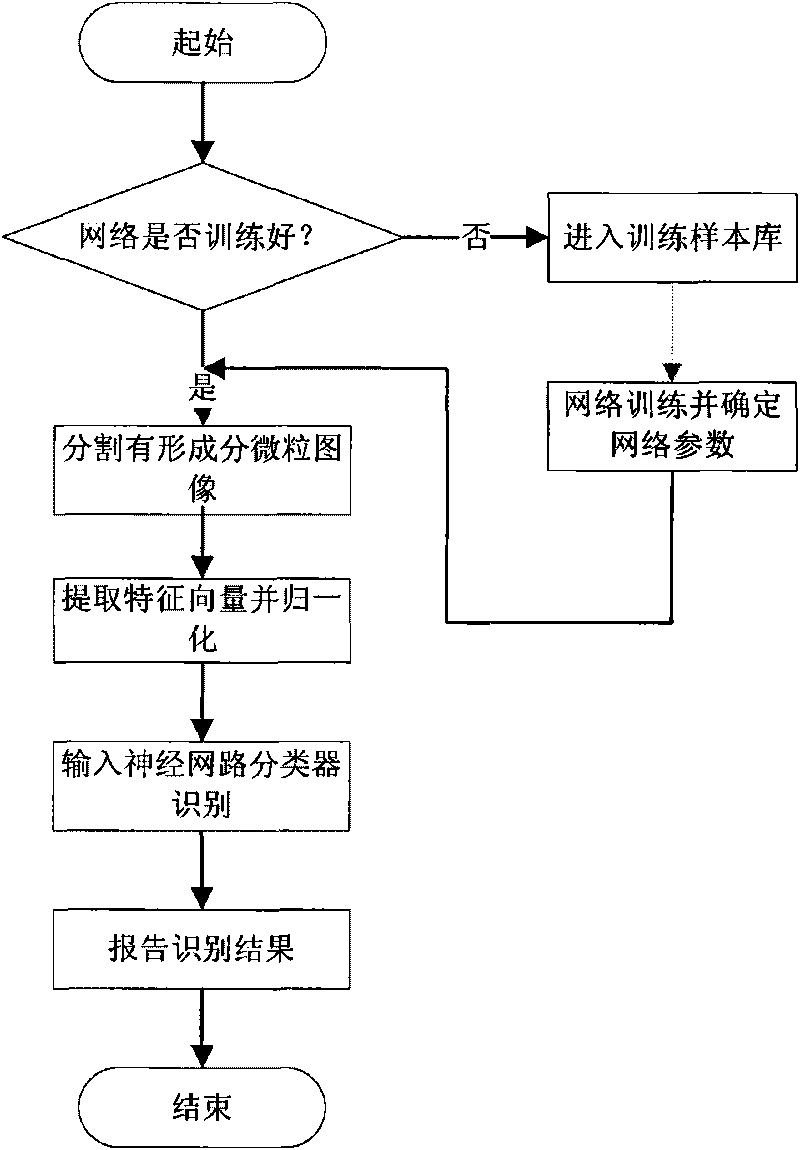
[0048] (1) Use the mobile microscope system in the urine sediment testing equipment to take pictures of urine samples, and then use the 1394 card to collect the images and transfer them to the memory of the computer of the urine sediment workstation. Here, each test sample needs to take 500 pictures .

[0049]...


PUM


Abstract

The invention relates to a neural network-based method for identifying and classifying visible components in urine, and belongs to a method for identifying and classifying the visible components in the urine. The method comprises the following steps: shooting an image of a urine sample with a flowing microscope system in urinary sediment detection equipment, and transmitting the image to a memoryof a urinary sediment image workstation; segmenting the shot image in the step 1 to form visible component particle images of the urine, calculating shape and texture feature vectors of the segmentedvisible component particle images in the step 2, and taking the vectors as input of an intelligent neural network; and receiving the feature vectors of the visible component particle images to be identified, normalizing to a range of [0,1], and inputting the trained intelligent neural network for identification. The method has high identification rate and low false positive rate, and greatly improves the accuracy and objectiveness of identifying the visible components in the clinical urine. Meanwhile, the workload of doctors is greatly lightened, and the standardization and automation of detecting the visible components in the urine are realized.

Description

technical field [0001] The invention belongs to the technical field of image processing, and in particular relates to a method for identifying and classifying urine formed components. Background technique [0002] Urine tangible analysis component detection is one of the three routine clinical inspection items. At present, there are three kinds of inspection and analysis methods commonly used in hospitals: microscope manual counting method, semi-automatic manual assisted identification analyzer and photoelectric signal method analyzer. [0003] Utilizing microscope manual counting method and semi-automatic manual assisted identification analyzer method to analyze and test the formed components of urine, the urine sample must first be centrifuged and pretreated, then photographed or observed, and finally manually identified. The recognition rate depends largely on the experience of the operator, and the efficiency is low. The photoelectric signal method analyzer is an instr...

Claims


Application Information

Patent Timeline
Patent Type & Authority Applications(China)
IPC IPC(8): G01N33/493G06N3/02G06N3/08G06T7/00
Inventor 宋洁沈继楠陈武
Owner DIRUI MEDICAL TECH CO LTD
Why Eureka
Try Eureka