Method for counting number of complex overlapping cells in microscopic image

A technology of microscopic image and statistical method, which is applied in the field of statistics of the number of complex cohesive cells in microscopic images, can solve the problems of seed point error, corrosion and expansion irreversible error, and difficult to find seed point for watershed segmentation, etc., to achieve small error , high precision effect

Inactive Publication Date: 2010-07-14
NINGBO UNIV
View PDF0 Cites 16 Cited by
  • Summary
  • Abstract
  • Description
  • Claims
  • Application Information

AI Technical Summary

Problems solved by technology

However, this method is relatively simple, and the times of corrosion and expansion must be manually determined empirically, and the irreversibility of corrosion and expansion will cause certain errors
Dimple segmentation is better for a small number of cells and the area

Method used


Image

  • Method for counting number of complex overlapping cells in microscopic image
  • Method for counting number of complex overlapping cells in microscopic image
  • Method for counting number of complex overlapping cells in microscopic image

Examples


Embodiment Construction

[0016] The present invention will be further described in detail below in conjunction with the accompanying drawings and embodiments.

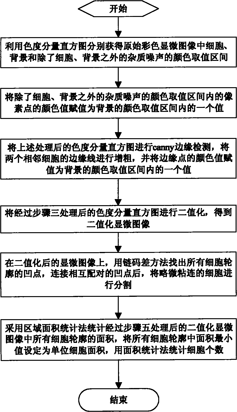
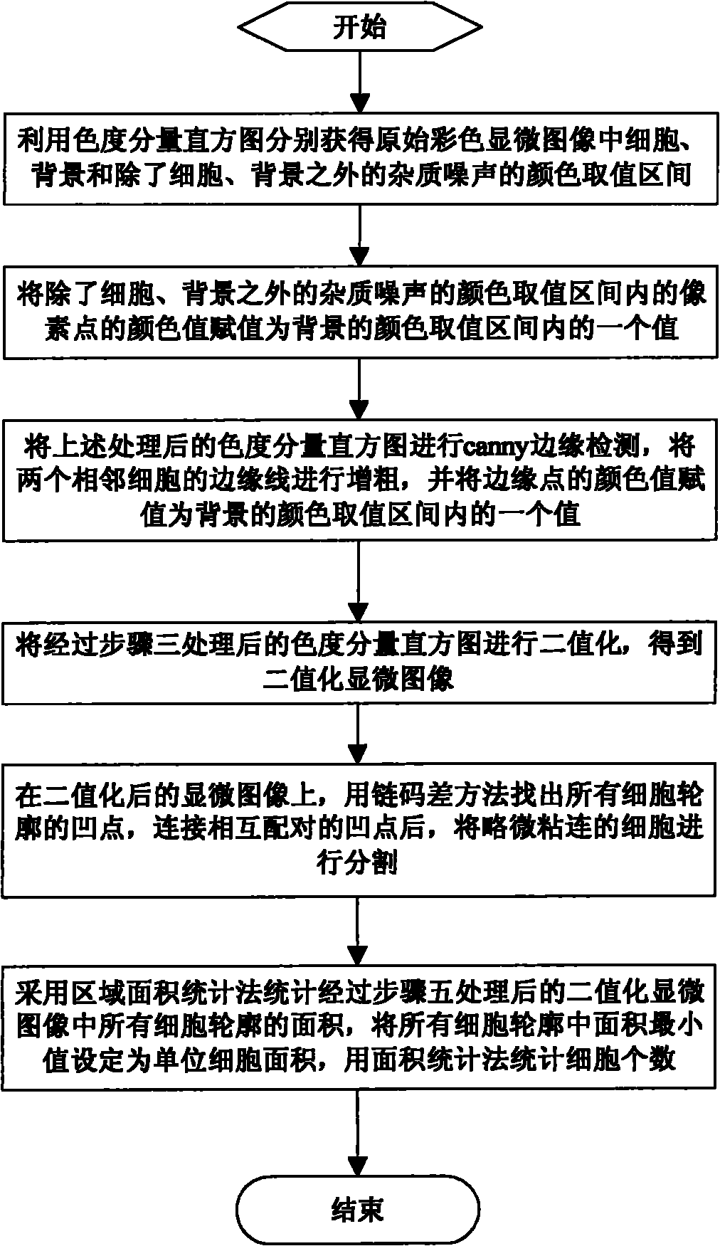
[0017] The invention provides a method for counting the number of complex adhesion cells of a microscopic image, which comprises the following steps: figure 1 as shown,

[0018] Step 1, using the chroma component histogram to respectively obtain the color value intervals of the cells, the background, and the impurity noise other than the cells and the background in the original color microscopic image;

[0019] Through the analysis of color microscopic images, especially blood cell images, it can be seen that chromaticity information plays a very important role in the discriminative analysis of cells. If only the color images are converted into grayscale images, it will undoubtedly lose some useful information for classification and judgment. information, so this step directly processes the color image; the chromaticity feature is the most wi...


PUM


Abstract

The invention relates to a method for counting the number of complex overlapping cells in a microscopic image. The method comprises the following steps: pre-processing and further binarizing an original colored microscopic image; locating the concave points in the outline of all the cells by using a chain code difference method, connecting the concave points which are paired with each other, and segmenting the slightly overlapping cells; and finally, counting the number of cells by using a region area statistical method. Compared with the prior art, the invention has the following advantages that: the original colored microscopic image is pre-processed and further binarized according to the color features of the colored microscopic image; the outline of the cells are detected and further segmented by using the chain code difference method because the slightly overlapping cells are characterized in that the concave points of the cells are obvious; and finally, the numbers of the heavily overlapping cells and the single cells are calculated by using the region area statistical method, so the method has small error and high accuracy.

Description

technical field [0001] The invention relates to the field of microscopic cell statistics, in particular to a method for counting the number of complicated adhesion cells in microscopic images. Background technique [0002] The microscopic cell image analysis includes the identification and measurement of the microscopic cell image, the statistics of the number, and the measurement of various parameters such as area, perimeter, and shape factor. In actual microscopic cell images, due to the slice and the cells themselves, multiple cells often overlap to form a larger area, such as blood cells, lymphocytes, cancer cells, etc. Counting the overlapping adherent cells that clump together is a difficult point in the analysis of microscopic cell images. At present, many segmentation algorithms have been proposed to deal with cell overlap and adhesion, such as erosion-dilation method based on mathematical morphology, approximate concave point cutting of binary image polygons, geode...

Claims


Application Information

Patent Timeline
IPC IPC(8): G06M11/00G06K9/62G06K9/38
Inventor 杨鸣杨翠翠蒋金涛邵华章湖王建宏
Owner NINGBO UNIV
Why Eureka
Try Eureka