Obtaining method of rat head magnetic resonance image Monte Carlo simulation model

A technology of magnetic resonance images and simulation models, applied in the field of image processing, can solve problems such as inability to obtain multi-tissue models, thin skulls, and small volumes, and achieve good practical application prospects and simplified complexity

Inactive Publication Date: 2010-09-29
NANJING UNIV OF AERONAUTICS & ASTRONAUTICS
View PDF3 Cites 8 Cited by
  • Summary
  • Abstract
  • Description
  • Claims
  • Application Information

AI Technical Summary

Problems solved by technology

Therefore, the segmentation of the five tissues cannot be completed by using threshold segmentation or fuzzy clustering directly through the gray information; due to the small size of the rat brain and the thin skull, ideal results cannot be obtained by using narrow-band level sets.
Therefore, the existing segmentation methods alone cannot obtain a multi-tissue model that meets the requirements of MC simulation.

Method used


Image

  • Obtaining method of rat head magnetic resonance image Monte Carlo simulation model
  • Obtaining method of rat head magnetic resonance image Monte Carlo simulation model
  • Obtaining method of rat head magnetic resonance image Monte Carlo simulation model

Examples


Embodiment Construction

[0025] The technical scheme of the present invention is described in detail below in conjunction with accompanying drawing:

[0026] as attached figure 1 Shown, the present invention is carried out according to the following steps:

[0027] A. Firstly, the image is divided into 6 parts including 5 tissues of scalp, skull, cerebrospinal fluid, gray matter and white matter, and image background;

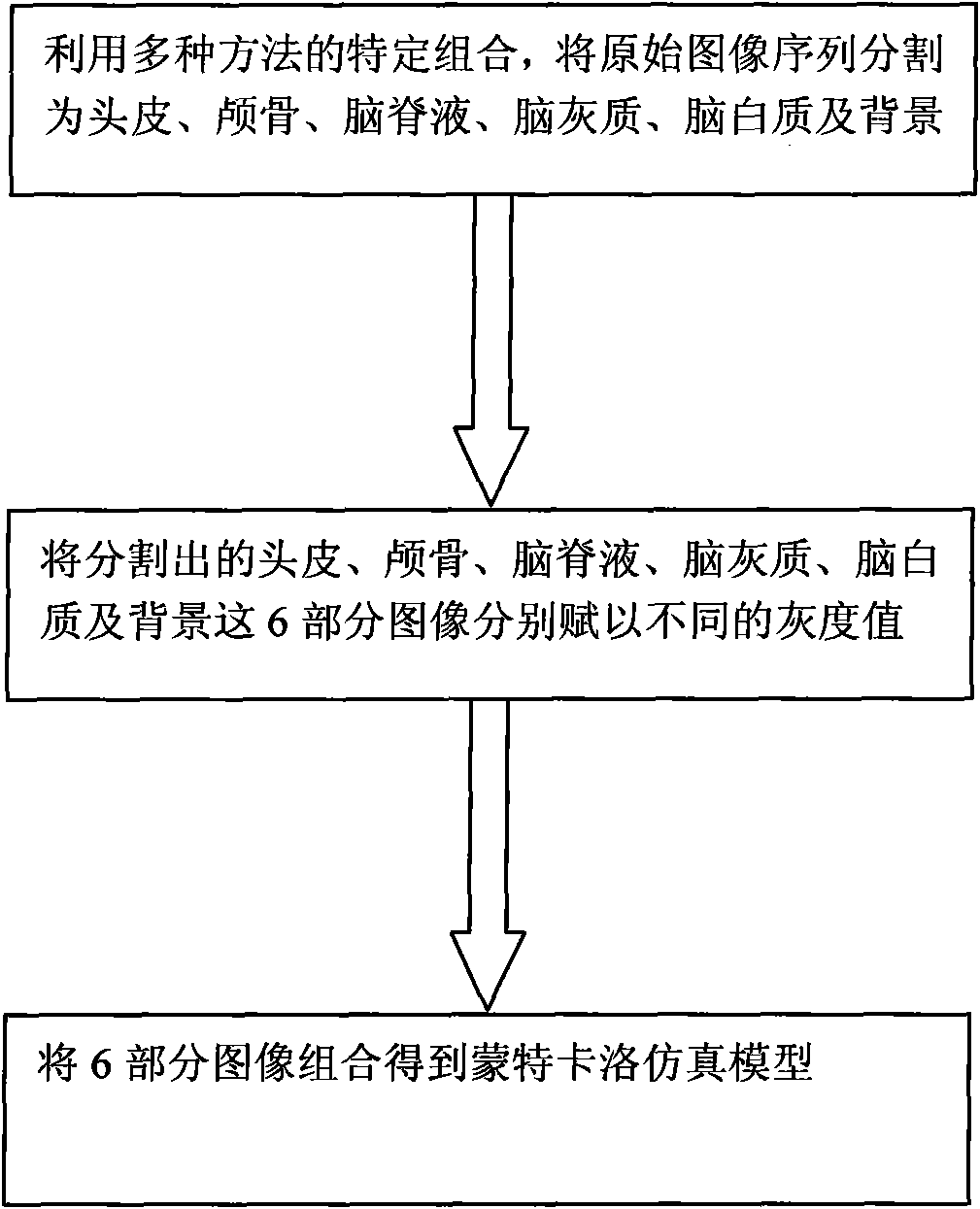
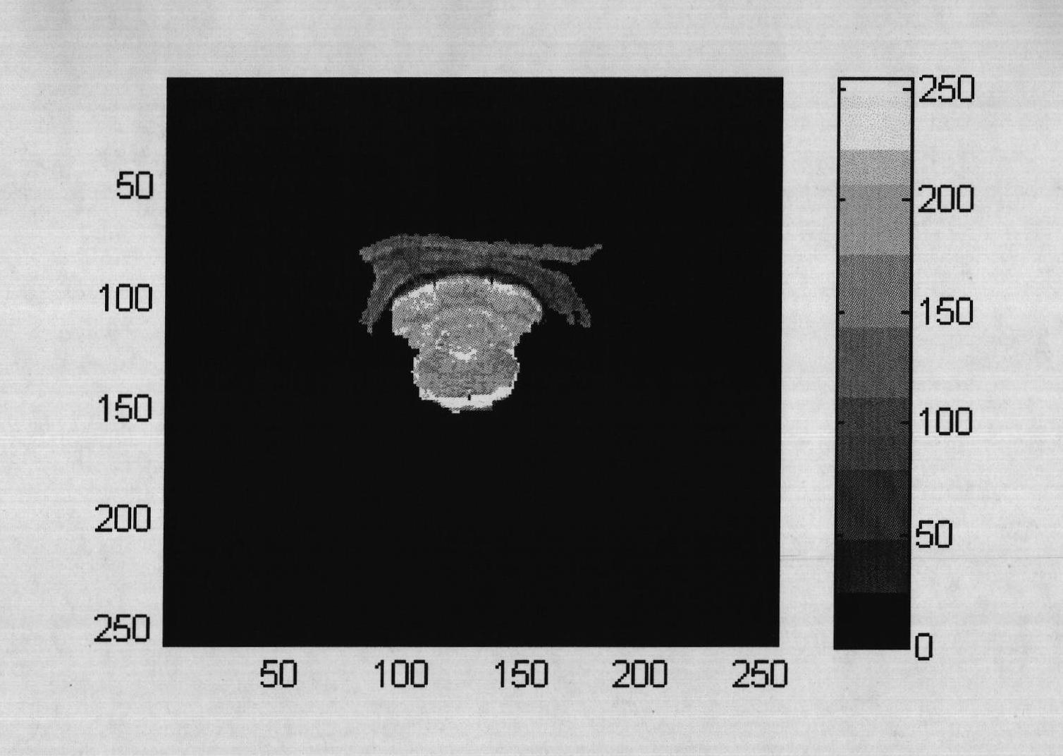
[0028] A1. Skull tissue segmentation: any image K0 in the original image sequence is processed using the grayscale threshold segmentation method (see attached figure 2 ) to segment, and use the expansion and erosion algorithm to obtain the skull tissue and discontinuous noise parts; then use the binary image maximum area marking method to remove the noise, that is, first mark the gray value of the discontinuous area of ​​the image, and then calculate the gray value of each marked area Area, where the continuous area with the largest area is the tissue to be segmented, its pixels are...


PUM


Abstract

The invention discloses an obtaining method of a rat head magnetic resonance image Monte Carlo simulation model, which belongs to the technical field of tissue optics. The obtaining method based on the specific combination of multiple image segmenting methods comprises the following steps of: firstly, determining a gray threshold value through analysis, and segmenting out skull tissues by adopting a threshold value segmentation, corrosion and expansion algorithm and a binary image maximum region labeling method; then removing all the tissues at the upper part of a skull in an original image, and segmenting out scalp tissues by adopting a narrow-band level set of special parameters; afterwards, segmenting out cerebrospinal fluid, ectocinerea and alba in the image in which the scalp and the skull are removed by adopting fast fuzzy clustering; and finally respectively endowing the five segmented tissues and background parts with different gray values to obtain a multi-tissue model. The invention can overcome the limitation of a single image segmenting method, rapidly and accurately obtains the rat head magnetic resonance image Monte Carlo simulation model, and has good practical application prospect.

Description

technical field [0001] The invention relates to an image processing method, in particular to a processing method for a rat head magnetic resonance image. Background technique [0002] By analyzing the incident light and the light scattered by the biological tissue, the optical parameter information inside the biological tissue can be obtained. The urgent problem to be solved is to study the propagation and distribution of light in the complex biological tissue. The Monte Carlo method (hereinafter referred to as MC) is a stochastic statistical method widely used to study the propagation and distribution of light in biological tissues. For the MC simulation of light distribution in heterogeneous complex tissues, the biological tissue must first be segmented according to different optical parameters (mainly: absorption coefficient, scattering coefficient, anisotropic factor) Different regions are formed to form a multi-tissue MC simulation model, and then the MC calculation is...

Claims


Application Information

Patent Timeline
Patent Type & Authority Applications(China)
IPC IPC(8): G06T7/00
Inventor 王新增李韪韬钱志余王雪娜
Owner NANJING UNIV OF AERONAUTICS & ASTRONAUTICS
Why Eureka
Try Eureka