Transesophageal echocardiography visual simulation system and method

A simulation system and cardiac technology, applied in the fields of virtual simulation and medical image analysis and processing, can solve problems such as difficult to further improve clinical operation assistance, unfavorable teaching and clinical data sharing, and failure to realize three-dimensional visualization section, so as to achieve good clinical simulation training Effect

Active Publication Date: 2012-07-04
WEST CHINA HOSPITAL SICHUAN UNIV +1
View PDF2 Cites 0 Cited by
  • Summary
  • Abstract
  • Description
  • Claims
  • Application Information

AI Technical Summary

Problems solved by technology

However, because the above-mentioned product belongs to a closed teaching system, clinical experience is not easy to be accumulated and expanded, which is not conducive to data sharing between teaching and clinical practice, and it is not easy to add clinical experience to the teaching system at any time
[0006] Japan's transesophageal ultrasound simulation teaching equipment was successfully developed by Japan's H R S Medical Systems Inc in 2008. This product includes analog probes, simulated people, and image databases, but it does not realize three-dimensional visualization, and the key to TEE learning is two-dimensional ultrasound. The embodiment of the cut surface on the 3D visualized heart is difficult to be further improved to clinical surgical assistance due to the lack of open modeling

Method used


Image

  • Transesophageal echocardiography visual simulation system and method
  • Transesophageal echocardiography visual simulation system and method
  • Transesophageal echocardiography visual simulation system and method

Examples


Embodiment 1

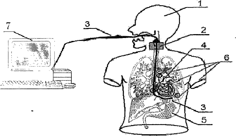
[0028] In this embodiment, the structure of the transesophageal echocardiography visualization simulation system is as follows: figure 1 As shown, it includes an intelligent phantom 1 , a cardiac ultrasound image simulation collection device 3 , a computer 7 connected to the cardiac ultrasound image simulation collection device, a transesophageal ultrasound probe posture data collection device 2 and a sensor 6 .

[0029] The smart phantom 1 includes a phantom and a three-dimensional simulated heart 4, a simulated esophagus and a stomach 5 arranged inside the phantom. The head and chest of the smart phantom are all made of transparent plastic, and the oral cavity of the head can be moved and opened and closed. , the head and neck can be separated, which can ensure the placement and removal of the transesophageal ultrasound probe attitude data acquisition device and sensors, and can directly observe the realistic visceral structure in the esophagus and chest cavity, and can see t...

Embodiment 2

[0037] This embodiment uses the transesophageal echocardiography visualization simulation system described in Example 1 to further illustrate the transesophageal echocardiography visualization simulation method. In this embodiment, the steps of the transesophageal echocardiography visualization simulation method are as follows:

[0038] The first step is to use the probe of the cardiac ultrasound image acquisition device and the transesophageal ultrasound probe attitude data acquisition device 2 to simultaneously acquire the probe attitude data of the transesophageal echocardiogram and the cardiac ultrasound image acquisition device in real time, and collect the acquired cardiac ultrasound images Establish a one-to-one correspondence relationship between the probe posture data of the device and the transesophageal echocardiogram, and store it in the database of the computer 7;

[0039]In the second step, on the basis of the standard virtual compound heart model (this model is ...


PUM


Abstract

The invention discloses a transesophageal echocardiography visual simulation system, comprising an intelligent phantom, a heart ultrasonic image stimulation and acquisition device, a computer connected with the heart ultrasonic image stimulation and acquisition device and a transesophageal ultrasonic probe attitude data acquisition device, wherein, the heart ultrasonic image stimulation and acquisition device is used for stimulating and acquiring heart ultrasonic images, and transmitting the acquired heart ultrasonic images to the computer; when the heat ultrasonic image stimulation and acquisition device acquires the heart ultrasonic images, the transesophageal echocardiography probe attitude data acquisition device and a sensor are used for synchronously acquiring probe attitude data ofthe heat ultrasonic image stimulation and acquisition device, and transmitting the acquired probe attitude data to the computer; and the computer is used for analyzing, processing and visually displaying the received heart ultrasonic images and the probe attitude data.

Description

technical field [0001] The invention belongs to the technical fields of medical image analysis and processing and virtual simulation, and in particular relates to a visual simulation system and method for transesophageal echocardiography teaching and training and computer-aided surgery. Background technique [0002] Ultrasound diagnosis of heart disease has always been the focus and difficulty in the field of ultrasound diagnosis. Transesophageal echocardiography is a new development in the field of cardiovascular disease diagnosis and monitoring, and provides a new means for the diagnosis of various heart diseases and large vessel diseases. The structure and movement of the heart are very complex, which puts higher demands on the examiner's spatial thinking ability and clinical thinking ability. Therefore, it is very important for doctors to quickly master echocardiography through high-quality training. There are many doctors and students at home and abroad who are in urgen...

Claims


Application Information

Patent Timeline
Patent Type & Authority Patents(China)
IPC IPC(8): G06F19/00G09B9/00
Inventor 宋海波姚宇赵向辉苗青李昕付忠良王伟东刘进
Owner WEST CHINA HOSPITAL SICHUAN UNIV
Why Eureka
Try Eureka