Medical appliance

A medical device and catheter technology, applied in the field of medical devices for the interventional diagnosis and treatment of gallbladder diseases, can solve problems such as resource waste, anastomotic stenosis, infection, etc., and achieve the effect of visual diagnosis and treatment

Active Publication Date: 2010-12-22
ANREI MEDICAL HZ
View PDF3 Cites 22 Cited by
  • Summary
  • Abstract
  • Description
  • Claims
  • Application Information

AI Technical Summary

Problems solved by technology

[0004] Clinically, this combination of laparoscopy and choledochoscopy for gallbladder preservation is often accompanied by complications such as postoperative infection, bile leakage, anastomotic stenosis, and peritonitis due to incisions

Method used


Image

  • Medical appliance
  • Medical appliance
  • Medical appliance

Examples


Embodiment 1

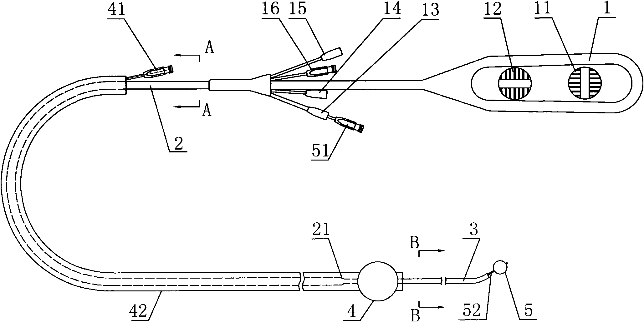
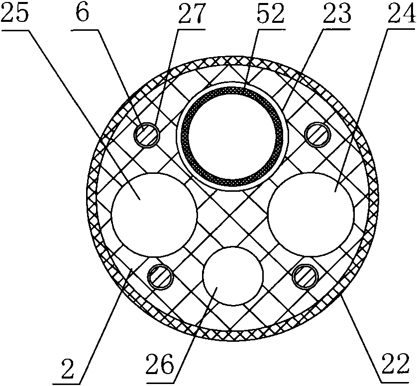
[0038] like Figure 1 to Figure 4 A medical device shown, including:

[0039] A catheter 2, the catheter 2 is provided with four main lumens 23, 24, 25, 26, and these lumens are independently connected from the proximal end of the catheter to the far end; the proximal end of the catheter 2 is provided with a Correspondingly communicated four plug-in ports 13, 14, 15, 16;

[0040] The first balloon 4, which can be inflated and deflated, is arranged at the distal end of the balloon sleeve 42, and the balloon sleeve 42 is movably sleeved outside the catheter 2, and the balloon The cannula 42 is provided with an air duct 43 capable of communicating with the first balloon 4 and the external balloon control device;

[0041]The second balloon 5, which can be inflated and deflated, is arranged at the distal end of the catheter 2, and the catheter 2 is provided with a ventilating channel capable of communicating with the second balloon 5 and the external balloon control device.

[0...

Embodiment 2

[0047] Such as Figure 5 The medical device described above is only different from Embodiment 1 in that the connection mode between the pulling wire and the terminal sheath is different: in this embodiment, the pulling wire 6 is fixedly sheathed on the metal at the distal end of the terminal sheath 3. Circle 7 is fixedly connected. The eyelet 7 is a steel ring. Others are the same as in Example 1.

Embodiment 3

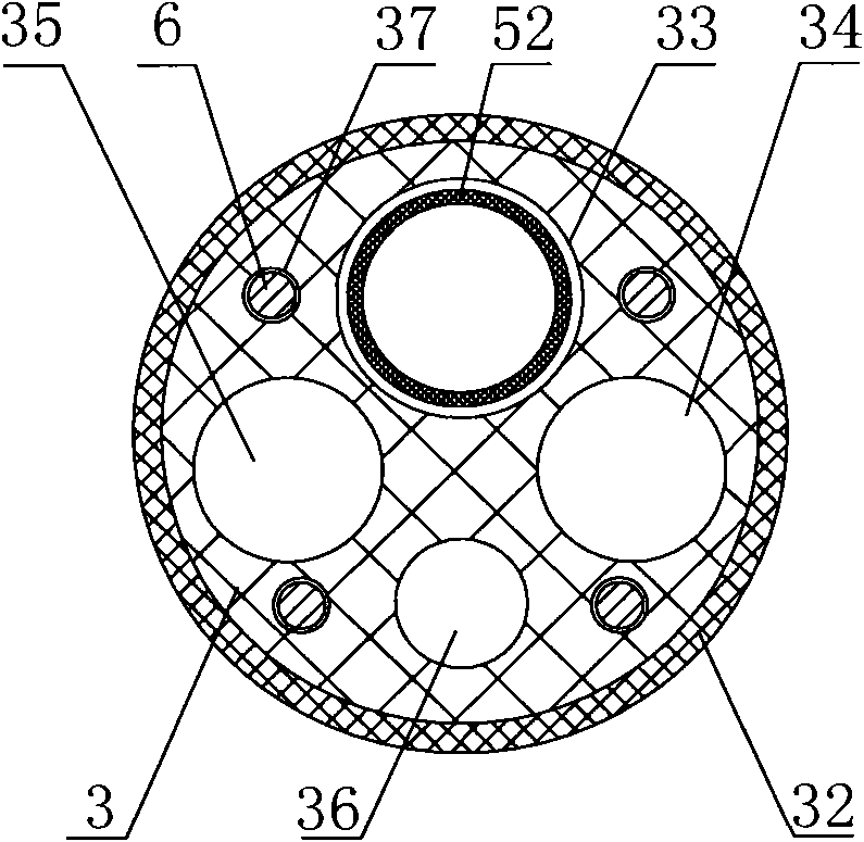
[0049] Such as Figure 6 to Figure 10 The medical device described above is only different from Embodiment 1 in that the ventilation duct of the second balloon is arranged differently: in this embodiment, the second balloon 5 is directly fixed on the end sheath tube 3, and the The ventilation channel of the second balloon 5 is formed by connecting the balloon lumen A29 on the catheter 2 and the balloon lumen B39 on the end sheath 3 , corresponding to the inflation port 19 of the balloon 5 . Others are the same as embodiment 1.

[0050] When the operation starts, the fiber optic mirror is inserted from the port 15, stretches out from the end of the terminal sheath tube 3, connects the fiber optic mirror with the imaging device and the light source, and guides the guide wire along the distal end of the terminal sheath tube 3. Enter the catheter 2, guide the terminal sheath 3 to be inserted into an endoscope (such as a duodenoscope), advance along the guide wire to the bifurcati...


PUM


Abstract

The invention discloses a medical appliance comprising a conduit, a first balloon and a second balloon, wherein the conduit is provided with at least two main cavity channels which perforates through the conduit from a near end to a far end, the near end of the conduit is provided with a plurality of insert ports which are correspondingly communicated with the cavity channels; the first balloon is capable of expanding after being inflated and contracting after being deflated, and is arranged at a far end of a balloon sleeve pipe, the balloon sleeve pipe is sleeved outside the conduit in a movable mode, and a ventilation pipeline which can be used for communicating the first balloon and an external balloon control device is arranged on the balloon sleeve pipe; and the second balloon is capable of expanding after being inflated and contracting after being deflated, and is arranged at the far end of the conduit, and a ventilation pipeline which can be used for communicating the second balloon and the external balloon control device is arranged on the conduit. The medical appliance can enter a gall bladder by using a double-balloon structure per se, can be provided with an imaging system to observe the gall bladder, and the jaw channel of the medical appliance per se can enter in other auxiliary appliances.

Description

technical field [0001] The invention relates to the technical field of gastroenterology medical devices, in particular to a medical device for interventional diagnosis and treatment of gallbladder diseases. Background technique [0002] Common gallbladder diseases include gallstones, cholecystitis, and gallbladder polyps. Due to the special location of the gallbladder physiological structure, the cystic duct and the opening of the cystic duct vary widely, and there are spiral mucosal folds in the cystic duct that need to be protected. Therefore, the treatment of gallbladder diseases generally adopts There are two ways of direct cholecystectomy and cholecystectomy. [0003] Traditional surgical treatment generally removes the gallbladder directly, but cholecystectomy will bring a variety of serious long-term side effects, which directly affect the quality of life of the patient, and even endanger the life of the patient. Therefore, with the advancement of science and technolo...

Claims


Application Information

Patent Timeline
IPC IPC(8): A61B19/00A61M25/10
Inventor 张婵娟张承
Owner ANREI MEDICAL HZ
Why Eureka
Try Eureka