Multi-mode molecular tomography system

An imaging system and molecular layer technology, applied in echo tomography, computerized tomography scanners, medical science, etc., can solve the problems of long reconstruction time, tomographic image fusion, and limited quality of reconstructed images, and achieve convenient collection and carrying effect

Inactive Publication Date: 2011-03-16
PEKING UNIV
View PDF4 Cites 28 Cited by
  • Summary
  • Abstract
  • Description
  • Claims
  • Application Information

AI Technical Summary

Problems solved by technology

Although fusion systems for tomographic (three-dimensional) images in XCT, FMT, and BLT modalities have also appeared, the fusion system follows the principle of first completing tomographic reconstruction for each modality, and then fusing each tomograp

Method used


Image

  • Multi-mode molecular tomography system
  • Multi-mode molecular tomography system
  • Multi-mode molecular tomography system

Examples


Embodiment Construction

[0028] The present invention will be described in detail below in conjunction with the accompanying drawings and embodiments.

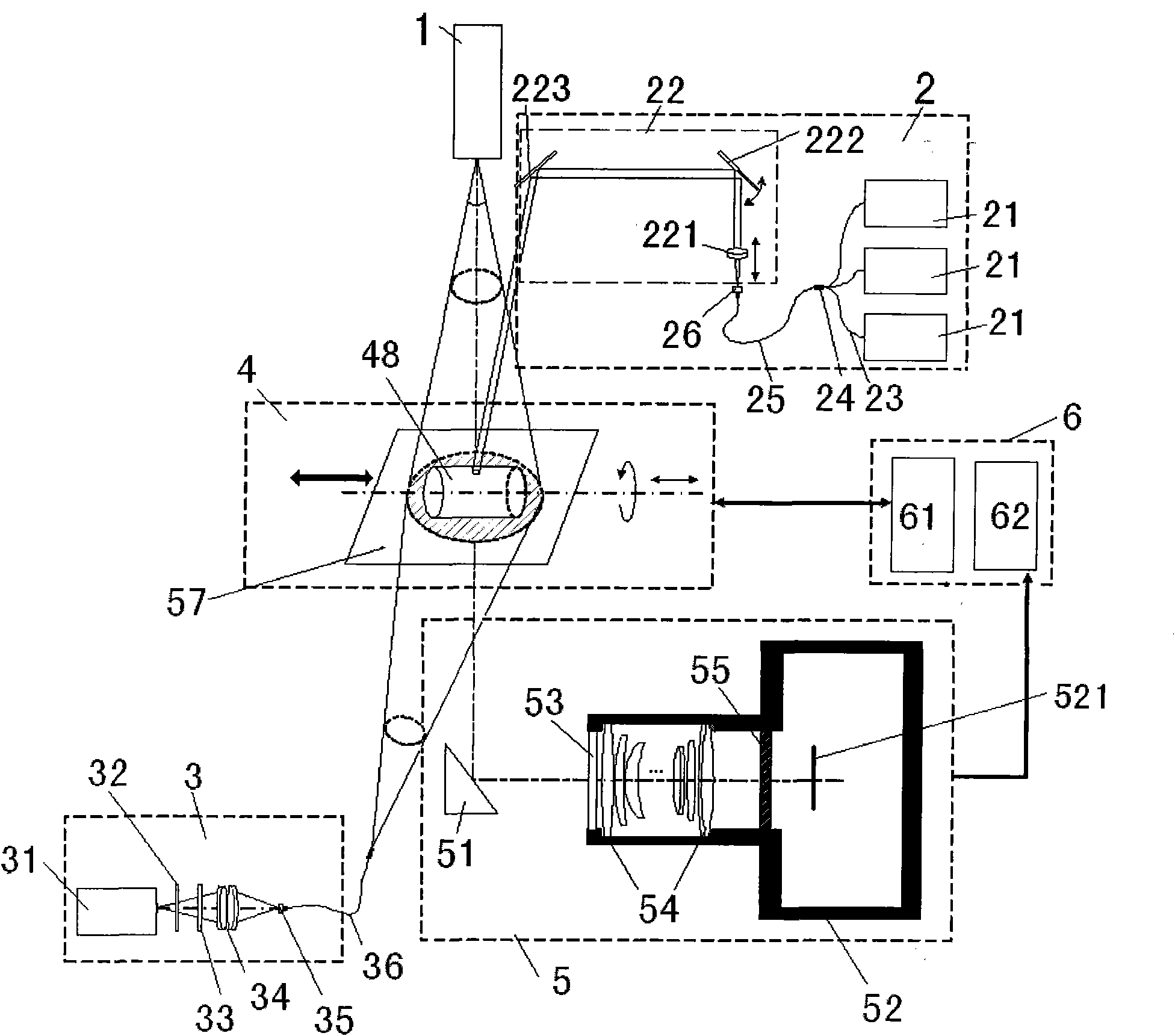
[0029] Such as figure 1 As shown, the present invention includes one or more of an X-ray source 1 , a near-infrared laser source 2 and a limited spectral width light source 3 , as well as a motorized object loading device 4 , an imaging device 5 and a control and processing device 6 . Under the control of the control and processing device 6, the motorized object loading device 4 drives the object to be scanned to translate and / or rotate 360° with the translation direction as the axis. One or more of them project scanning light onto the object to be scanned. The imaging device 5 measures and photoelectrically converts the light intensity distribution emitted from the surface of the object to be scanned after being scanned by the light source. The control and processing device 6 obtains the light intensity distribution emitted from the surface of the ...


PUM


Abstract

The invention relates to a multi-mode molecular tomography system which is characterized by comprising one or more light sources of an X-ray source, a near infrared laser light source and a finite spectral width light source for projecting scanning light to an object to be scanned, an electric loading device, an imaging device and a control and processing device, wherein the imaging device is used for obtaining intensity distribution data of x-rays, visible light or near infrared light emerging from the surface of the object to be scanned after scanning, and inputting the intensity distribution data into the control and processing device; the control and processing device is used for controlling the actions of the object to be scanned through the electric loading device; and the control and processing device comprises a tomography module, the tomography module is used for receiving the data of the imaging device, utilizing XCT (X-ray computed tomography) and DOT (diffuse optical tomography) modes to reconstruct an outer boundary and similar information of all internal organizations during marginalization, fusing and reconstructing XCT, DOT, FMT (fluorescence molecular tomography) and BLT (bioluminescence tomography) single-mode or multi-mode tomography image after fusion and outputting. The multi-mode molecular tomography system is applicable to the field of x-ray and optical biomedical imaging.

Description

technical field [0001] The invention relates to a molecular tomography imaging system, in particular to a multi-mode molecular tomography imaging system. Background technique [0002] Currently, computer tomography (CT, Computed Tomography) imaging technology can be used to realize non-invasive three-dimensional imaging of living organisms, including structural tomography and functional tomography of living organisms. Among them, structural tomography techniques include X-ray tomography (XCT), nuclear magnetic resonance imaging (MRI, nuclear magnetic resonance imaging), positron emission tomography (PET, Positron Emission Tomography) and single photon emission tomography (SPECT, single photon emission computed tomography), etc., have been widely used in clinical diagnosis, but the above-mentioned various imaging techniques are only aimed at imaging the internal structural features of a certain type of organisms, and most of the systems are expensive. Optical molecular-based...

Claims


Application Information

Patent Timeline
IPC IPC(8): A61B19/00A61B5/00A61B6/03A61B90/00
Inventor 毛珩杨建生卢彦斌司远征周宇约翰·华莱士·埃默森周铁姜明
Owner PEKING UNIV
Why Eureka
Try Eureka