
Quantitative multi-spectral opto-acoustic tomography (MSOT) of tissue biomarkers

A technology of tissue biology and photoacoustic tomography, applied in the direction of using tomography for diagnosis, diagnosis, diagnostic recording/measurement, etc., can solve the problem of difficult realization of the illumination field, and achieve the effect of excellent spatial resolution

Active Publication Date: 2011-07-27
健康与环境慕尼黑德国研究中心赫姆霍茨中心(有限公司)
View PDF11 Cites 50 Cited by
  • Summary
  • Abstract
  • Description
  • Claims
  • Application Information

AI Technical Summary

Problems solved by technology

Such an illumination field is difficult to achieve in practice due to the unknown tissue heterogeneity and the ability to impose significant changes in light intensity on any given plane within the tissue

Method used


Image

  • Quantitative multi-spectral opto-acoustic tomography (MSOT) of tissue biomarkers
  • Quantitative multi-spectral opto-acoustic tomography (MSOT) of tissue biomarkers
  • Quantitative multi-spectral opto-acoustic tomography (MSOT) of tissue biomarkers

Examples


Embodiment Construction

[0068] Now referring specifically to the drawings in detail, it is emphasized that the details are shown by way of example and for the purpose of illustrative discussion of only the preferred embodiments of the present invention, and in order to provide the principles and principles of the present invention considered to be the most useful and easy to understand. The conceptual description presents these details. In this regard, no attempt is made to show the structural details of the present invention in more detail than necessary for a basic understanding of the present invention. Figure one It will be obvious to those skilled in the art how several forms of the invention can be embodied in practice. As used herein, unless explicitly stated to exclude, elements or steps recited in the singular and beginning with the word "a" or "an" should be understood as not excluding multiple elements or steps. In the description of the drawings, similar numbers represent similar parts. ...


PUM


Abstract

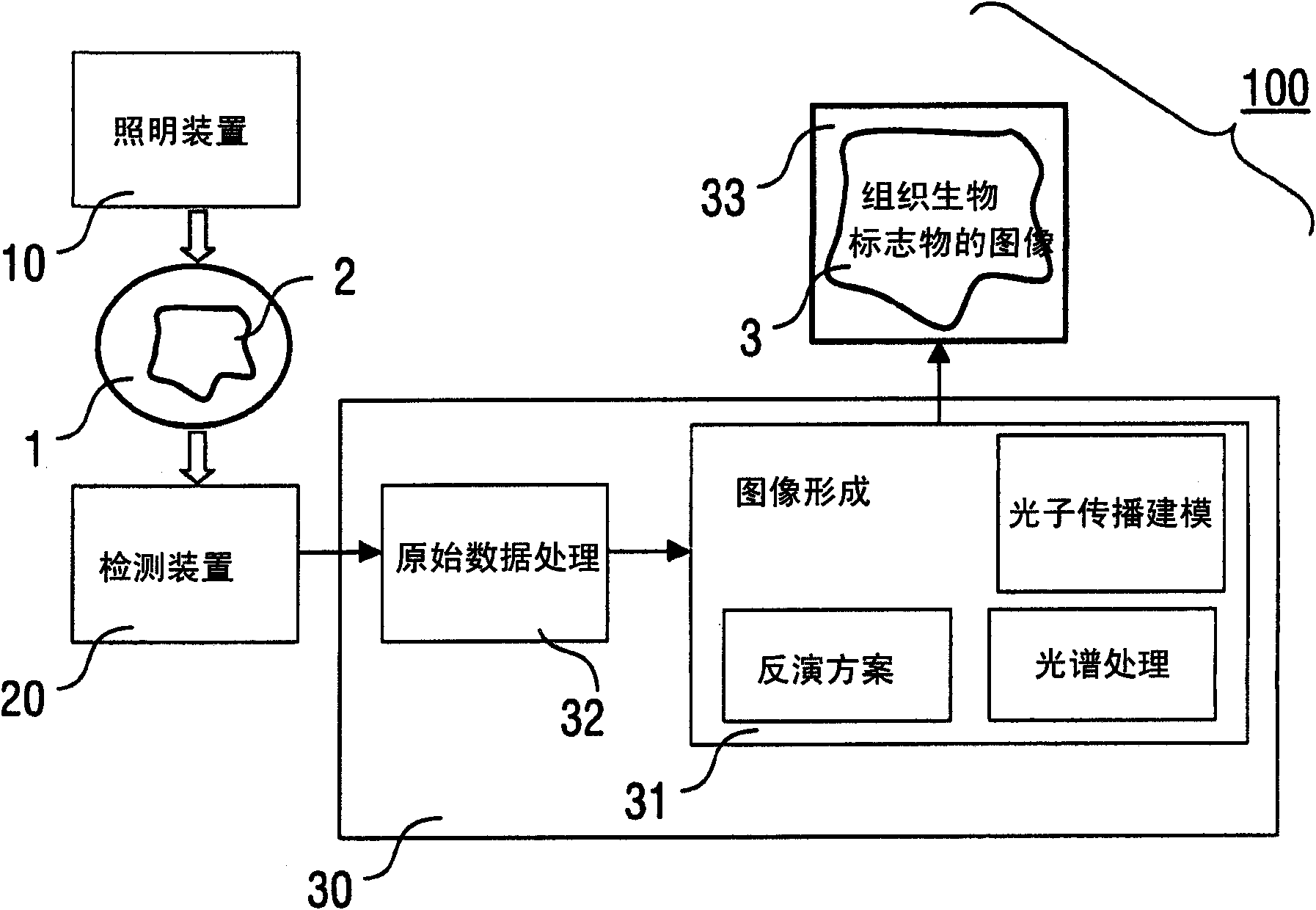
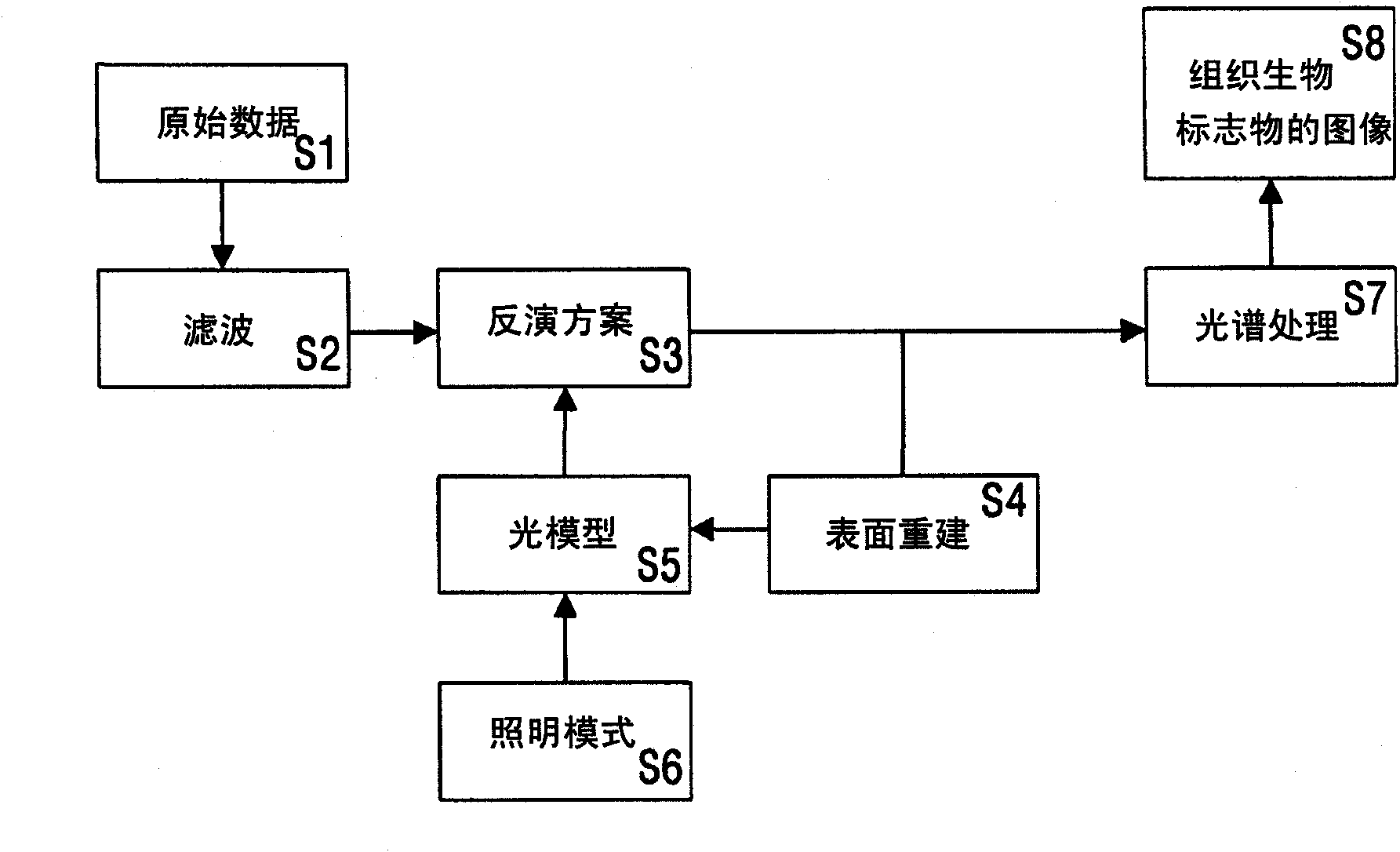
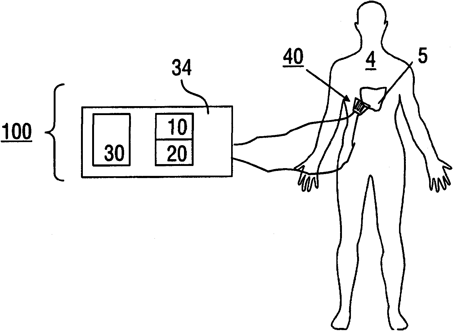
A method of multi-spectral opto-acoustic tomography (MSOT) imaging of a target tissue including a target tissue biomarker comprises the steps of illuminating the target tissue with an illumination device emitting at least one pulsed illumination pattern at several illumination wavelengths, detecting pressure signals from the target tissue biomarker with a detector device, wherein the pressure signals being produced in the target tissue in response to the said illumination, and reconstructing a quantitative tomographic image of a distribution of the target tissue biomarker in the target tissue, wherein the pressure signals are analyzed using a photon propagation model, which depends on an illuminating light fluence in the target tissue and on the illumination wavelengths, at least one spectral processing scheme, and an inversion scheme providing the tomographic image. Furthermore, an imaging device for multi-spectral opto-acoustic tomography is described.

Description

Technical field [0001] The present invention relates to methods and devices for quantitative three-dimensional sensing and imaging of target tissue markers, which are especially used in clinical, small animal and small biological imaging applications using multi-wavelength illumination. Background technique [0002] Non-invasive imaging of in vivo functions and molecular markers is an emerging and important capability in biological discovery, drug discovery, and some clinical applications, which goes beyond anatomical imaging and delayed disease identification. Another important prospect of visualizing tissue biomarkers is the ability to detect and quantify the treatment response in vivo by monitoring specific major molecules or downstream targets. The therapeutic effect can then be dynamically detected on the time scale from hour to day. This ability is in contrast to current mainstream medical care with traditional late-stage end-point efficacy, and this practice often comprom...

Claims


Application Information

Patent Timeline
Patent Type & Authority Applications(China)
IPC IPC(8): A61B5/00
CPCA61B5/0095A61B5/0073
Inventor D・拉扎斯基V・恩齐亚赫里斯托斯
Owner 健康与环境慕尼黑德国研究中心赫姆霍茨中心(有限公司)
Learn More