Shape appliance digitalizing method of cleft palate alveolar bone

A technology of alveolar bone and appliances, applied in the medical field, can solve the problem that the accuracy cannot meet the digital precision design of alveolar bone shaping appliances

Inactive Publication Date: 2011-08-31
FOURTH MILITARY MEDICAL UNIVERSITY
View PDF2 Cites 11 Cited by
  • Summary
  • Abstract
  • Description
  • Claims
  • Application Information

AI Technical Summary

Problems solved by technology

Existing CT includes ordinary tomographic CT and spiral CT, which belong to macroscopic CT at the level of naked eye resolution. Due to the limitation of its technical principle, the scanning slice thickness is generally 5-10mm. Even if ultra-thin sl

Method used


Image

  • Shape appliance digitalizing method of cleft palate alveolar bone
  • Shape appliance digitalizing method of cleft palate alveolar bone
  • Shape appliance digitalizing method of cleft palate alveolar bone

Examples


Embodiment Construction

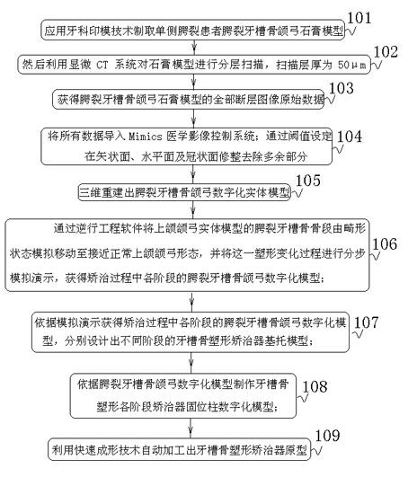
[0030] Such as figure 1 As shown, a digital method for cleft palate alveolar bone shaping appliance, at least includes the following steps:

[0031] Step 101, using dental impression technology to make a plaster model of the alveolar bone and jaw arch of patients with unilateral cleft palate;

[0032] Step 102, and then use the micro-CT system to scan the plaster model in layers, and the scanning layer thickness is 50 μm;

[0033] Step 103, obtaining the original data of all tomographic images of the plaster model of the cleft palate alveolar bone and maxillary arch;

[0034] Step 104, importing all data into the Mimics medical image control system; trimming and removing redundant parts on the sagittal plane, horizontal plane and coronal plane through threshold setting;

[0035] Step 105, three-dimensional reconstruction of the digital solid model of the cleft palate alveolar bone and jaw arch;

[0036] In step 106, the cleft palate alveolar bone segment of the maxillary ar...


PUM

PropertyMeasurementUnit
Diameteraaaaaaaaaa
Lengthaaaaaaaaaa

Abstract

The invention belongs to a non-invasive treating method of congenital cleft palate in the medical field, especially relating to a shape appliance digitalizing method of a cleft palate alveolar bone, which comprises the following steps of: 1) obtaining the jaw arch fault information of a cleft palate alveolar bone by using the microscopic CT (computed tomography) technology, wherein the thickness of a scanning layer is 50 mu m; 2) moving the cleft palate alveolar bone segment of a maxillary arch solid model to approach to a normal maxillary arch form from an abnormal form in a simulation manner in reverse engineering software, and simulating and demonstrating the shape change process step by step; 3) obtaining a digitalized model of the cleft palate alveolar bone jaw arch in each stage in the process of remedying according to the simulation and demonstration; 4) manufacturing an appliance retention column digitalized model used in each stage of shaping the alveolar bone according to the digitalized model of the cleft palate alveolar bone jaw arch; and 5) automatically processing a prototype of an alveolar bone shape appliance by using the rapid forming technology. A digitalized designing and manufacturing method of the shape appliance of a cleft palate alveolar bone independent of the experience and handwork of a doctor is provided.

Description

technical field [0001] The invention belongs to the non-traumatic treatment method for congenital cleft palate in the medical field, and in particular relates to a digital method for a cleft palate alveolar bone shaping appliance. Background technique [0002] Cleft lip and palate is the most common congenital maxillofacial defect deformity in the world. In China, the incidence of cleft palate is about 1.6‰ of the birth population, which brings great burden to society and family. Cleft palate directly leads to severe maxillary alveolar bone tissue defect in the patient, resulting in the loss of continuity of the maxillary arch and tissue displacement of the alveolar bone segment, which brings many difficulties to subsequent operations, so alveolar bone plastic surgery is required. A large number of clinical studies have proved that the maxillary alveolar bone of infants with cleft palate has strong plasticity. The use of appliances for alveolar bone shaping can maximize the ...

Claims


Application Information

Patent Timeline
IPC IPC(8): G06F17/50
Inventor 吴国锋魏建华甘云娜谢诚马威
Owner FOURTH MILITARY MEDICAL UNIVERSITY
Why Eureka
Try Eureka