
Method for detecting P53 protein expression in brain tumor

A P53 protein and detection method technology, applied in special data processing applications, instruments, electrical digital data processing, etc., can solve problems such as inability to guide the formulation of preoperative treatment plans, subjective influence of test results, etc., to avoid subjective influence, Avoid the effects of insufficient standardization and low cost

Inactive Publication Date: 2011-09-28
ZHEJIANG UNIV
View PDF3 Cites 7 Cited by
  • Summary
  • Abstract
  • Description
  • Claims
  • Application Information

AI Technical Summary

Problems solved by technology

[0003] Currently, the most widely used clinical detection method for P53 protein is immunohistochemical technique, which requires surgery to obtain pathological sections of glioma before detection, so it cannot guide the formulation of preoperative treatment plan
At the same time, research by Zhou Xiaojun (Zhou Xiaojun. The correct application of immunohistochemistry in pathological diagnosis. Journal of Diagnostic Pathology. 2003, 10(4): 232-235) showed that immunohistochemical techniques have deficiencies in standardization and quantification of results. The results are susceptible to the subjective influence of the inspector
Currently there is no method for P53 protein detection based on image processing and pattern recognition technology

Method used


Image

  • Method for detecting P53 protein expression in brain tumor
  • Method for detecting P53 protein expression in brain tumor
  • Method for detecting P53 protein expression in brain tumor

Examples


Embodiment Construction

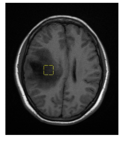
[0015] The brain tumor P53 protein expression detection method based on magnetic resonance image analysis of the present invention comprises the following steps:

[0016] (1) Acquiring magnetic resonance images of brain tumor patients, wherein the magnetic resonance images include any one or more of T1-weighted sequences, T1-enhanced sequences, and FLAIR sequences. The specific collection method is as follows:

[0017] A magnetic resonance scanner (such as GE Healthcare, 1.5T) is used to acquire magnetic resonance images in the transverse, coronal or sagittal positions of glioma patients, and the magnetic resonance images include T1-weighted sequences, T1-enhanced sequences and FLAIR sequences. Among them, the imaging parameters of T1 weighted sequence are preferably Repetition Time=1966.1ms, Echo Time=21.088ms, Inversion Time=750ms; the imaging parameters of T1 enhanced sequence are preferably Repetition Time=1967.25ms, Echo Time=7.264ms, Inversion Time= 750ms; the imaging p...


PUM


Abstract

The invention discloses a magnetic resonance image analysis-based method for detecting P53 protein expression in a brain tumor. The method comprises the following steps: (1) collecting magnetic resonance images of a T1 weighted sequence, a T1 reinforced sequence and a fluid attenuated inversion recovery (FLAIR) sequence of a patient with brain tumor; (2) intercepting lesion area images in the magnetic resonance image from anyone of the sequences, combining the lesion area images into a lesion area image set, and marking the lesion area images as P53 protein expression positive or negative; (3) analyzing images of the lesion area image set, extracting image characteristics in the lesion area images, and combining the image characteristics into a lesion area sample set; (4) randomly selecting part of samples from the lesion area sample set as a training sample set, and combining other samples into a verification sample set, and training a classifier by using the training sample set; and(5) classifying the verification samples by using the trained classifier to obtain the situation of the brain tumor P53 protein expression of the verification samples.

Description

technical field [0001] The invention belongs to the technical field of image processing and pattern recognition, and in particular relates to a method for detecting the expression status of brain tumor P53 protein through magnetic resonance image analysis. Background technique [0002] Glioma is the most common tumor of the central nervous system. It has the characteristics of long treatment cycle, easy recurrence, high morbidity and mortality, and poses a great threat to the health and life of patients. At present, early diagnosis of glioma can be achieved, and timely surgery, radiotherapy and chemotherapy can be given, but the prognosis of patients with malignant glioma has not been significantly improved. Afshar et al. (Golnar Afshar, Nannette Jelluma, Yang Xiaodong et al. Radiation-Induced Caspase-8Mediates p53-Independent Apoptosis in Glioma Cells. Cancer Research. 2006, 66(8): 4223-4232) showed that p53 in adult glioma patients Gene mutations are associated with impro...

Claims


Application Information

Patent Timeline
IPC IPC(8): G06F19/18G06F19/24G06K9/62
Inventor 夏顺仁刘晨彬潘颖
Owner ZHEJIANG UNIV
Learn More