Brain tumor MIB-1 index range detection method
A MIB-1, detection method technology, applied in the fields of instruments, character and pattern recognition, computer parts, etc., can solve the problem of insufficient standardization of immunohistochemical techniques and quantification of results, and the detection results are susceptible to the subjective influence of the testing personnel, and cannot be guided. Formulate preoperative treatment plans and other issues to achieve the effect of avoiding subjective influence, avoiding insufficient standardization and low cost
- Summary
- Abstract
- Description
- Claims
- Application Information
AI Technical Summary
Problems solved by technology
Method used
Image
Examples
Embodiment 1
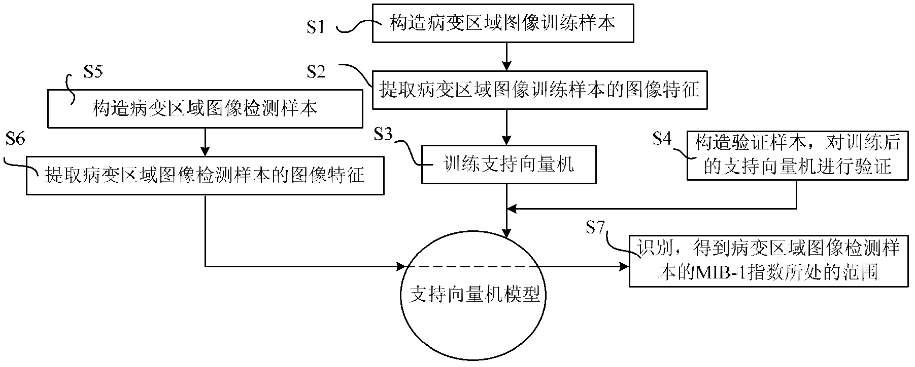
[0046] Such as figure 1 A method for detecting the range of brain tumor MIB-1 index includes the following steps:
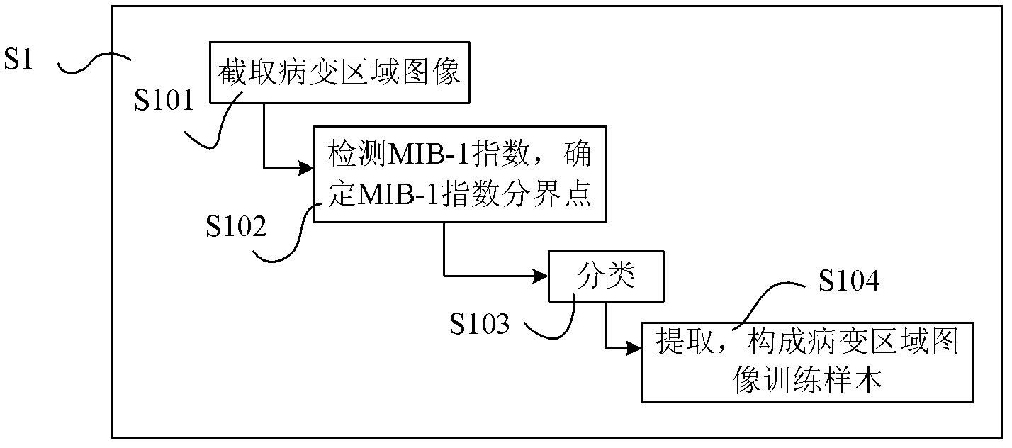
[0047] S1 collects magnetic resonance images of brain tumor patients and constructs image training samples of lesion regions;
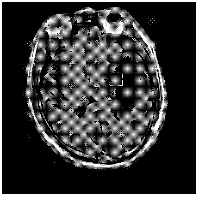
[0048] In this step, the magnetic resonance images include any one or more of T1-weighted sequences, T1-enhanced sequences, and FLAIR sequences, and the specific acquisition methods are as follows:
[0049] A magnetic resonance scanner (such as GE Healthcare, 1.5T) is used to acquire magnetic resonance images in the transverse, coronal or sagittal positions of glioma patients, and the magnetic resonance images include T1-weighted sequences, T1-enhanced sequences and FLAIR sequences. Among them, the imaging parameters of T1 weighted sequence are preferably Repetition Time=1966.1ms, Echo Time=21.088ms, Inversion Time=750ms; the imaging parameters of T1 enhanced sequence are preferably Repetition Time=1967.25ms, EchoTime=7.264ms, Inversion...
Embodiment 2
[0246] Due to the high dimensionality of the image feature sample set of the lesion area, it may contain some redundant image features. On the one hand, these redundant image features may reduce the classification accuracy, and on the other hand, it will greatly increase the computational overhead of the support vector machine. ; Therefore, as a preference, the lesion area image feature sample set can be optimized based on the discrete particle swarm optimization algorithm to obtain the lesion area image optimization feature set; determine the parameters of the support vector machine model according to the lesion area image optimization feature set, effectively reduce the image feature of complexity.
[0247] In particle swarm optimization, each potential solution to an optimization problem can be imagined as a point in the search space, called a particle. The quality of the particle's current position is evaluated by the objective function, and the objective function calculat...
PUM
 Abstract
Description
Claims
Application Information
 - R&D
- Intellectual Property
- Life Sciences
- Materials
- Tech Scout
- Unparalleled Data Quality
- Higher Quality Content
- 60% Fewer Hallucinations
Browse by: Latest US Patents, China's latest patents, Technical Efficacy Thesaurus, Application Domain, Technology Topic, Popular Technical Reports.
© 2025 PatSnap. All rights reserved.Legal|Privacy policy|Modern Slavery Act Transparency Statement|Sitemap|About US| Contact US: help@patsnap.com


