
Brain tumor MIB-1 index range detection method

A MIB-1, detection method technology, applied in the fields of instruments, character and pattern recognition, computer parts, etc., can solve the problem of insufficient standardization of immunohistochemical techniques and quantification of results, and the detection results are susceptible to the subjective influence of the testing personnel, and cannot be guided. Formulate preoperative treatment plans and other issues to avoid subjective influences, avoid insufficient standardization, and reduce costs

Inactive Publication Date: 2013-04-24
ZHEJIANG UNIV
View PDF4 Cites 0 Cited by
  • Summary
  • Abstract
  • Description
  • Claims
  • Application Information

AI Technical Summary

Problems solved by technology

[0003] In the prior art, the most widely used clinical detection method for MIB-1 index is immunohistochemical technique, which needs surgery to obtain the patient's glioma pathological section before detection, which causes great trauma to the patient and cannot guide Develop a preoperative treatment plan
An article "The Correct Application of Immunohistochemistry in Pathological Diagnosis" published on pages 232-235 of "Journal of Diagnostic Pathology" in 2003 showed that immunohistochemical techniques have deficiencies in standardization and quantification of results, and the test results are vulnerable to The subjective influence of the tester
At present, there is no method for MIB-1 index range detection based on image processing and pattern recognition technology

Method used


Image

  • Brain tumor MIB-1 index range detection method
  • Brain tumor MIB-1 index range detection method
  • Brain tumor MIB-1 index range detection method

Examples


Embodiment 1

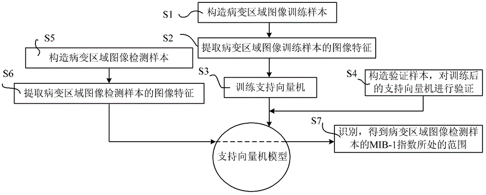
[0046] like figure 1 A brain tumor MIB-1 index range detection method is shown, including the following steps:

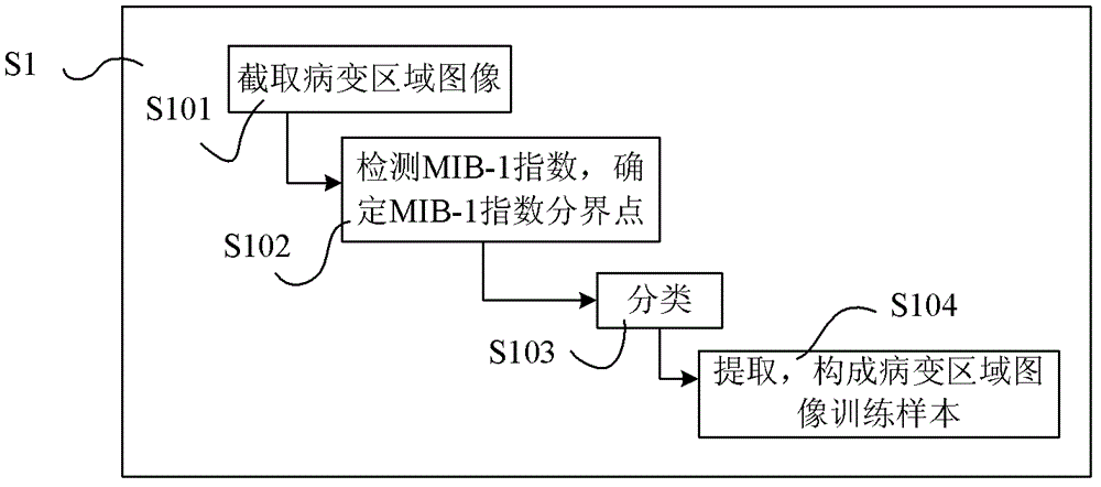
[0047] S1 collects magnetic resonance images of brain tumor patients, and constructs image training samples of lesion areas;

[0048] In this step, the magnetic resonance image includes any one or several of the T1 weighted sequence, the T1 enhanced sequence, and the FLAIR sequence, and the specific acquisition method is as follows:

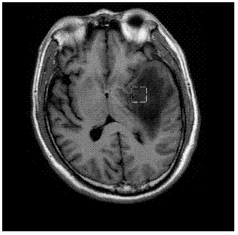
[0049] Transverse, coronal, or sagittal magnetic resonance images of glioma patients are acquired using a magnetic resonance scanner (eg, GE Healthcare, 1.5T), including T1-weighted, T1-enhanced, and FLAIR sequences. Among them, the imaging parameters of the T1 weighted sequence are preferably Repetition Time=1966.1ms, Echo Time=21.088ms, Inversion Time=750ms; the imaging parameters of the T1 enhanced sequence are preferably Repetition Time=1967.25ms, EchoTime=7.264ms, Inversion Time=750ms ; The imaging parameters of the FLAIR sequen...

Embodiment 2

[0246] Due to the high dimensionality of the image feature sample set in the lesion area, it may contain some redundant image features. On the one hand, these redundant image features may reduce the classification accuracy, and on the other hand, will greatly increase the computational cost of the support vector machine. Therefore, as an option, the image feature sample set of the lesion area can be optimized based on the discrete particle swarm algorithm to obtain an optimized image feature set of the lesion area; the parameters of the support vector machine model are determined according to the optimized feature set of the lesion area image, which can effectively reduce the image features. complexity.

[0247] In particle swarm optimization, each potential solution to an optimization problem can be imagined as a point in the search space, called a particle. The quality of the particle's current position is evaluated by the objective function, and the objective function calcu...


PUM


Abstract

The invention relates to a detection method for obtaining a brain tumor MIB-1 index range by analyzing a magnetic resonance image of a patient with brain tumor. The method comprises the following steps of: acquiring the magnetic resonance image of the patient with brain tumor, and forming a lesion area image training sample; extracting image features of the lesion area image training sample, training to obtain a support vector machine model according to the extracted image features of the lesion area image training sample; forming a lesion area image detection sample, and extracting the imagefeatures of the lesion area image detection sample; detecting and obtaining the range of the MIB-1 index of the lesion area image detection sample by the support vector machine model according to theimage features of the lesion area image detection sample. The method solves the defect that the MIB-1 index only can be detected by immune tissue chemical detection after obtaining the brain tumor pathological tissue in operation in the prior art, and avoids the problems of introduction of subjective thought of detectors and poor standardization in the immune tissue chemical detection.

Description

technical field [0001] The invention relates to image processing and identification technology, in particular to a detection method for obtaining the range of brain tumor MIB-1 index by analyzing magnetic resonance images of brain tumor patients. Background technique [0002] Gliomas are the most common tumors of the central nervous system, accounting for approximately 42% of primary brain tumors. Although with the development of medical technology, the treatment of glioma has gradually developed into a comprehensive therapy based on surgery, combined with radiotherapy, chemotherapy, immunotherapy, etc., but the prognosis of many patients has not improved significantly. Clinically, phosphatase and tensin homolog deleted onchromosome 10 (PTEN), epidermal growth factor receptor (EGFR), theO6-methylguanine-DNA methyltransferase (MGMT), tumor protein 53 (P53), monoclonal antigen antibody of cell proliferation associated nuclear (MIB- 1) and other protein expression status, and ...

Claims


Application Information

Patent Timeline
Patent Type & Authority Patents(China)
IPC IPC(8): G06K9/62G06K9/46
Inventor 夏顺仁潘颖刘晨彬
Owner ZHEJIANG UNIV
Learn More