
Signal detection method and device based on multi-chip ultrasonic sensors

An ultrasonic sensor and signal detection technology, which is applied in the field of biomedical signal processing, can solve the problems of signal deterioration, difficult operation, and high risk of infection, and achieve accurate intermediate data processing, increase ultrasonic output, and reduce burden. Effect

Active Publication Date: 2012-06-20
EDAN INSTR
View PDF3 Cites 15 Cited by
  • Summary
  • Abstract
  • Description
  • Claims
  • Application Information

AI Technical Summary

Problems solved by technology

[0003]In actual clinical application, the fetus will turn over or swim in the amniotic fluid of the mother (that is, commonly known as fetal movement in the industry), and the probe is in a fixed position. After the fetus moves, If the fetal heart rate is not in the ultrasound coverage area of ​​the probe, the signal will deteriorate, which will affect the quality of monitoring. It is necessary for the medical staff to adjust the position of the probe in time and align the fetal heart rate again; more commonly, in the second stage of labor, that is, during the delivery process, the The position of the fetal heart rate keeps changing and the position of the probe is fixed, which will inevitably lead to poor signal captured by the probe and poor monitoring effect. Now there are two commonly used solutions. One is that the medical staff constantly repositions the fetal heart rate and Adjust the position of the probe in time to regain a better signal, but this increases the burden on the medical staff and increases the difficulty of monitoring; the second method is to use direct fetal electrocardiography (Direct ECG), with a spiral electrode hooked to the scalp of the fetus In general, the fetal heart rate is calculated by using the fetal ECG signal, but this method is invasive, difficult to operate, and has a high risk of infection

Method used


Image

  • Signal detection method and device based on multi-chip ultrasonic sensors
  • Signal detection method and device based on multi-chip ultrasonic sensors
  • Signal detection method and device based on multi-chip ultrasonic sensors

Examples


Embodiment Construction

[0048] In order to make the object, technical solution and advantages of the present invention more clear, the present invention will be further described in detail below in conjunction with the accompanying drawings and embodiments. It should be understood that the specific embodiments described here are only used to explain the present invention, not to limit the present invention.

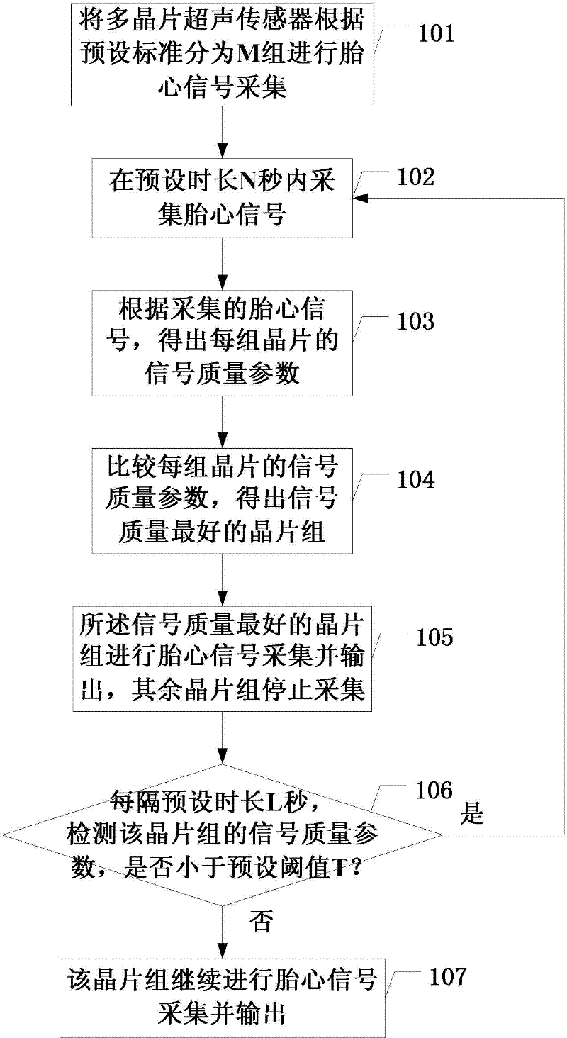
[0049] Such as figure 1 Shown, the specific steps of a kind of signal detection method based on multi-chip ultrasonic sensor of the present invention are:

[0050] 101. Divide the multi-chip ultrasonic sensors into M groups according to preset standards for fetal heart signal acquisition;

[0051] The ultrasonic sensor is composed of multiple wafers. According to the preset standard, the present invention preferably divides the multiple wafers into those with the same number, equal distances between adjacent points, forming a symmetrical pattern structure, and shared wafers between adjacent gr...


PUM


Abstract

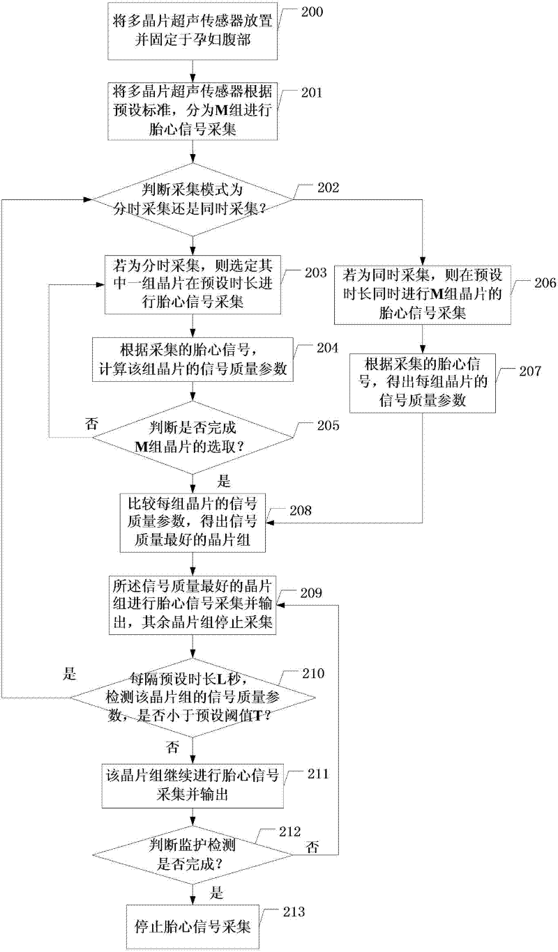
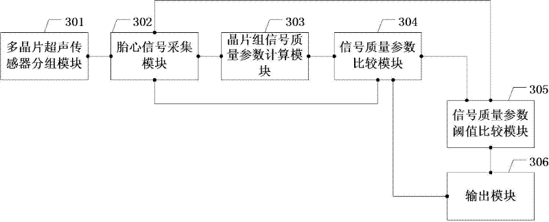
The invention relates to the field of biomedical signal processing, in particular to a signal detection method and device based on multi-chip ultrasonic sensors. The method comprises the following steps: dividing the multi-chip ultrasonic sensors into M groups according to the preset standard; carrying out fetal heart signal acquisition in preset duration; obtaining the signal quality parameter of each chip group according to the acquired fetal heart signal; obtaining the chip group with the best signal quality through comparison; carrying out fetal heart signal acquisition and output on the chip group and stopping acquisition of other chip groups; detecting whether the signal quality parameter of the chip group is less than the preset threshold every preset duration, if so, repeating theabove steps, and if no, continuing fetal heart signal acquisition and output. The method and the device have the following beneficial effects: the burdens of the medical workers are lightened; more accurate intermediate processing data are obtained; meanwhile, the sound field combined by the chips is uniform; and only a group of chips work in a combined manner in the normal custody process, thus neither increasing power consumption nor increasing ultrasonic output quantity.

Description

technical field [0001] The invention relates to the field of biomedical signal processing, in particular to a signal detection method and device based on a multi-chip ultrasonic sensor. Background technique [0002] At present, the ultrasonic Doppler fetal monitor, which is widely used in the field of obstetrics, utilizes a characteristic of sound - the Doppler principle, which means that the sound will shift in frequency when it meets the echo reflected by a moving object. Proportional to the speed of the moving object. The ultrasound probe of the fetal monitor is fixed on a certain position in the abdomen of the pregnant woman with a strap (usually the area that can be well aligned with the fetal heart), emits ultrasound toward the fetal heart, and receives echoes. When the valve velocity is within a certain range, the monitoring equipment can collect the echo data reflected by the fetal heart after amplifying and filtering processing, and also use this echo cycle data to...

Claims


Application Information

Patent Timeline
IPC IPC(8): A61B5/0444A61B5/344
Inventor 陈德伟曾永华汪培林张雷
Owner EDAN INSTR
Learn More