Method for constructing three-dimensional brain model
A brain model, three-dimensional technology, which is applied in the field of building 3D digital brain models to ensure the quality of medical care and improve the efficiency of teaching
- Summary
- Abstract
- Description
- Claims
- Application Information
AI Technical Summary
Problems solved by technology
Method used
Image
Examples
Embodiment 1
[0043] step 1
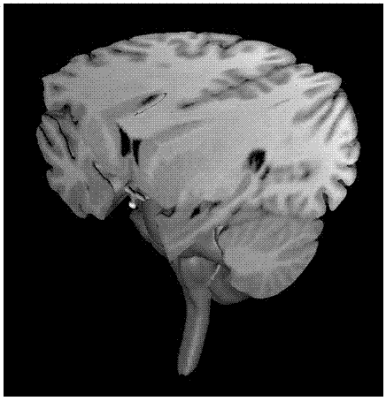
[0044] A 34-year-old healthy male volunteer, 1.5T magnetic resonance head sagittal thin layer imaging, the obtained T 1 The original data in DICOM format are the materials of this study. Imaging conditions: layer thickness 0.8mm, layer distance 0.95mm; W×H=256×256; number of layers 170.
[0045] Preprocess the magnetic resonance image into bmp format.
[0046] step 2
[0047] Graphics workstation: Core central processing unit, memory, color display, 1024 × 768 resolution, storage unit; Windows XP operating system, 3DS-MaX11.0, mimics8.0 and related software.
[0048]Separate the target image of the brain, select the bmp format image of the human part of the brain, import it into the mimics software, import the preprocessed bmp format image into the specified layer in sequence, and set the spatial orientation of the image through the spatial orientation setting module , so that the spatial position of the brain in the picture set by the spatial orientation s...
Embodiment 2
[0060] step 1
[0061] 34-year-old healthy male volunteer, 1.5T magnetic resonance head sagittal thin-section imaging, obtained T 1 The raw data in DICOM format is the data for this study. Imaging conditions: layer thickness 0.8mm, layer spacing 0.95mm; W×H=256×256; the number of layers is 170.
[0062] The magnetic resonance images are preprocessed into bmp format.
[0063] Step 2
[0064] Graphics workstation: Core CPU, memory, color display, 1024 × 768 resolution, storage unit; Windows XP operating system, 3DS-Max11.0, mimics8.0 and related software.
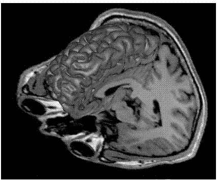
[0065] Select the magnetic resonance image of the part of the human brain after removing the part described in Example 1, import the mimics software, import the bmp format image obtained by preprocessing into the specified layer in order, set the spatial orientation of the picture, and make the image in the picture. The spatial position of the brain set by the spatial orientation setting module corresponds to the actual s...
Embodiment 3
[0077] step 1
[0078] 34-year-old healthy male volunteer, 1.5T magnetic resonance head sagittal thin-section imaging, obtained T 1 The raw data in DICOM format is the data for this study. Imaging conditions: layer thickness 0.8mm, layer spacing 0.95mm; W×H=256×256; the number of layers is 170.
[0079] The magnetic resonance images are preprocessed into bmp format.
[0080] Step 2
[0081] Graphics workstation: Core CPU, memory, color display, 1024 × 768 resolution, storage unit; Windows XP operating system, 3DS-Max11.0, mimics8.0 and related software.
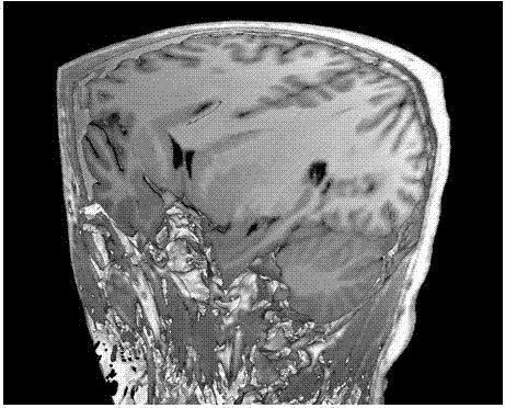
[0082] Selected from the magnetic resonance images of the part of the human brain described in Example 1 and the part including the skull surrounding the part, imported into the mimics software, and the bmp format images obtained by preprocessing are sequentially imported into the designated layers, and the spatial orientation setting module is used. Set the spatial orientation of the picture, so that the spatial position...
PUM
| Property | Measurement | Unit |
|---|---|---|
| Layer thickness | aaaaa | aaaaa |
| Layer thickness | aaaaa | aaaaa |
Abstract
Description
Claims
Application Information
 - R&D
- Intellectual Property
- Life Sciences
- Materials
- Tech Scout
- Unparalleled Data Quality
- Higher Quality Content
- 60% Fewer Hallucinations
Browse by: Latest US Patents, China's latest patents, Technical Efficacy Thesaurus, Application Domain, Technology Topic, Popular Technical Reports.
© 2025 PatSnap. All rights reserved.Legal|Privacy policy|Modern Slavery Act Transparency Statement|Sitemap|About US| Contact US: help@patsnap.com


