Microwave induced thermoacoustic tomography system for early discovery and diagnosis of breast cancer

A technology for early detection and thermal-induced ultrasound, applied in diagnosis, sonic diagnosis, infrasound diagnosis, etc., can solve the problems of low imaging resolution, large volume, ionization hazards, etc., and achieve high imaging resolution, high imaging resolution, and detection. Sensitive effect

Active Publication Date: 2012-10-10
UNIV OF ELECTRONICS SCI & TECH OF CHINA
View PDF10 Cites 8 Cited by
  • Summary
  • Abstract
  • Description
  • Claims
  • Application Information

AI Technical Summary

Problems solved by technology

The above two detection methods require large-scale equipment detection equipment as support, large volume, and low imaging resolution, which makes do

Method used


Image

  • Microwave induced thermoacoustic tomography system for early discovery and diagnosis of breast cancer
  • Microwave induced thermoacoustic tomography system for early discovery and diagnosis of breast cancer
  • Microwave induced thermoacoustic tomography system for early discovery and diagnosis of breast cancer

Examples


Embodiment 1

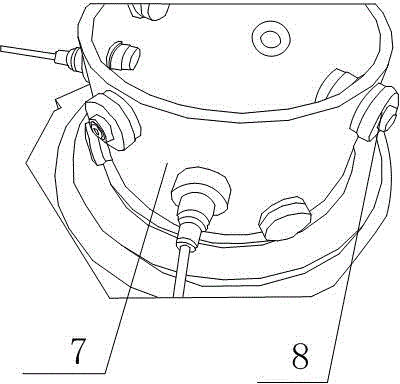
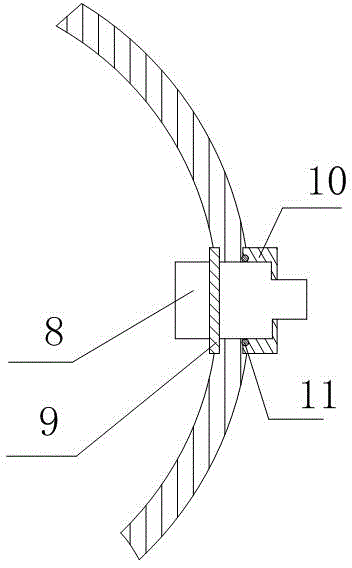
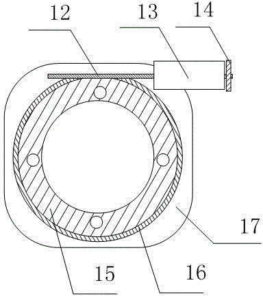
[0030] Such as figure 1 As shown, the microwave thermal ultrasound imaging system for early detection and diagnosis of breast cancer includes a circular scanning turntable 17, a radiation antenna is arranged inside the circular scanning turntable 17, a scanning bowl 7 is arranged above the radiation antenna, and the scanning bowl 7 is set Above the circular scanning turntable 17 , several ultrasonic probes 8 are arranged on the scanning bowl 7 ; several preamplifiers are arranged below the circular scanning turntable 17 , and the preamplifiers are connected to the ultrasonic probes 8 . In the conversion process of electromagnetic energy to acoustic energy, the electromagnetic energy is radiated into the space by the antenna and penetrates the tissue under test, and the ultrasonic signal is emitted by the diseased tissue and propagated to the ultrasonic probe 8 to be converted back into an electrical signal. During this process, the peak power of the electromagnetic wave energy...

Embodiment 2

[0038] Such as Figure 6 , Figure 7 , Figure 8 As shown, the structure of embodiment 2 is basically the same as that of embodiment 1, except that the preamplifier is mainly composed of AD797, fourth-order filter, OPA847, fourth-order filter, and OPA846 connected in sequence.

[0039] The ultrasonic signal is weak (the peak-to-peak value is around 200uV). In order to realize the sampling of the signal and the digital signal processing in the later stage, the signal needs to be amplified first. Since the dynamic range of the input signal of the data acquisition card is ±2V, in order to make full use of the resolution of the acquisition card, it is expected to amplify the signal to this range, so the voltage gain of the amplifier is about 4V / 200uV=20000. The spectral width of the ultrasonic pulse signal is about 50KHz 800KHz, but in order to achieve high-fidelity effects on ultrasonic signals, the bandwidth of the amplifier should be much greater than 800KHz. Since multipl...


PUM


Abstract

The invention discloses a microwave induced thermoacoustic tomography system for early discovery and diagnosis of breast cancer. The system comprises a circular scanning turntable, a radiating antenna is arranged in the circular scanning turntable, a scanning bowl is arranged above the radiating antenna, the scanning bowl is arranged above the circular scanning turntable, and a plurality of ultrasonic probes are arranged on the scanning bowl; and a plurality of preamplifiers are arranged below the circular scanning turntable and connected with the ultrasonic probes. Through the system, tumor tissues are converted into an ultrasonic pulse source under electromagnetic wave pulse irradiation by using strong contrast of the tumor tissues and normal tissues on electromagnetic energy absorption and the advantage of high short wavelength imaging resolution of ultrasonic and using a microwave induced thermoacoustic effect, and the tumor tissues are imaged and positioned by using a time reversal imaging technology, so that ionizing radiation harm is not produced, and the detection sensitivity and the imaging resolution of the system are higher than those of the conventional B ultrasonic equipment; and the system tends to frontier research and equipment development in the field of tumor detection.

Description

technical field [0001] The invention relates to a microwave thermal ultrasonic imaging system, which is mainly used for early detection and diagnosis of breast cancer. Background technique [0002] Breast cancer is one of the most common malignant tumors in women. According to statistics, the incidence rate accounts for 7-10% of all kinds of malignant tumors in the whole body. Its incidence is often related to heredity, and the incidence rate is higher in women between the ages of 40 and 60 and before and after menopause. Only about 1-2% of breast cancer patients are men. Malignant tumors usually occurring in the glandular epithelium of the breast. Breast cancer is one of the most common malignant tumors that seriously affects women's physical and mental health and even threatens their lives. Male breast cancer is rare. There are two traditional detection methods, one is to use mammography, which is a non-invasive method. The Siemens-ElemaAB type mammography machine is u...

Claims


Application Information

Patent Timeline
IPC IPC(8): A61B8/00
Inventor 赵志钦朱晓章柳清伙聂在平刘昱朗牛骏吴江牛宋健王金国
Owner UNIV OF ELECTRONICS SCI & TECH OF CHINA
Why Eureka
Try Eureka