Method and device for processing ultrasonic images and breast cancer diagnosis equipment

A technology of ultrasound images and diagnostic equipment, applied in character and pattern recognition, instruments, computer parts, etc., can solve the problems of inaccurate histogram, inability to solve the problem of irregular tumor detection, inaccuracy, etc.

Inactive Publication Date: 2013-01-02
BEIJING SAMSUNG TELECOM R&D CENT +1
View PDF6 Cites 14 Cited by
  • Summary
  • Abstract
  • Description
  • Claims
  • Application Information

AI Technical Summary

Problems solved by technology

The breast computer-aided detection system (B-CAD) developed by Canada's Medipattern can reduce the time it takes for radiologists to analyze breast ultrasound images. It can automatically analyze, segment and classify images, but still requires manual input to achieve highly accurate results.
[0004] In addition, there are some methods using a single feature and a single classifier, but these methods cannot solve the problem of irregular tumor detection
For example, US7736313 relates to the detection of les...

Method used


Image

  • Method and device for processing ultrasonic images and breast cancer diagnosis equipment
  • Method and device for processing ultrasonic images and breast cancer diagnosis equipment
  • Method and device for processing ultrasonic images and breast cancer diagnosis equipment

Examples


Embodiment Construction

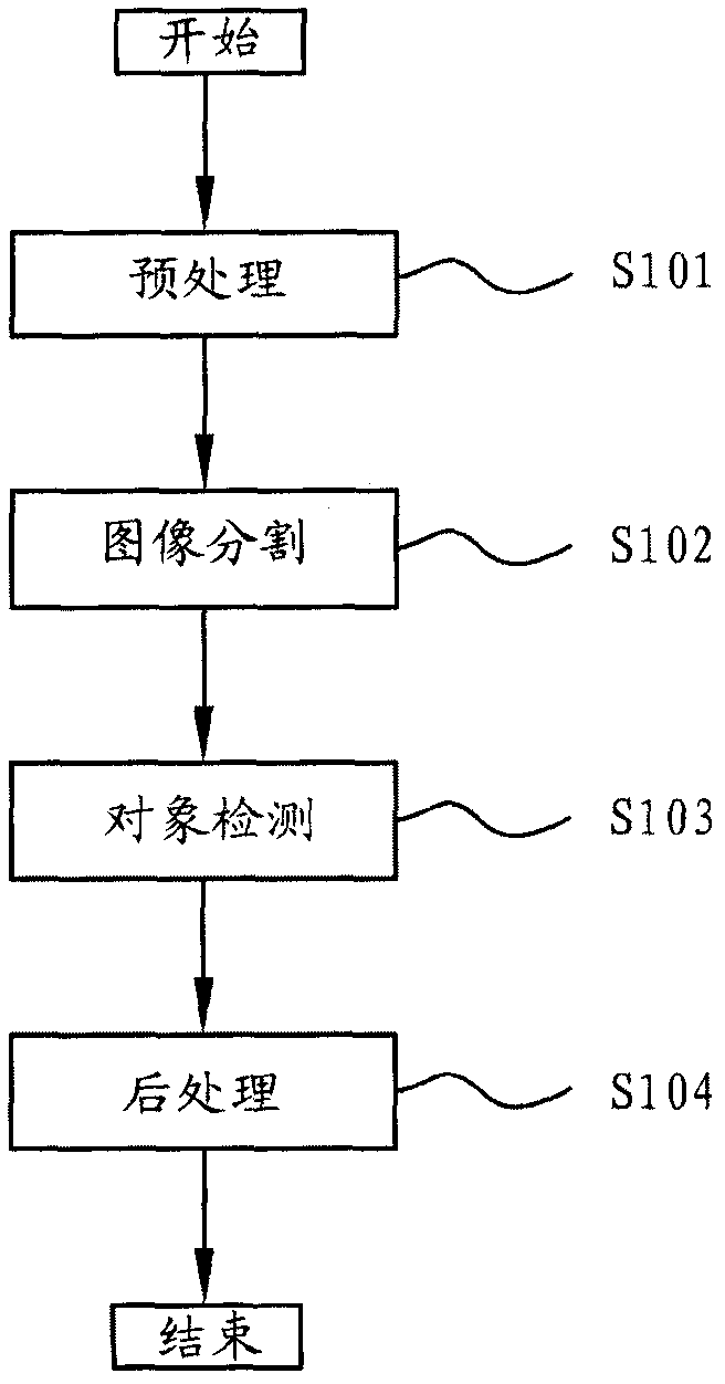
[0037] figure 1 is a flowchart illustrating a method of processing an ultrasound image according to the present invention. Such as figure 1 As shown, the method for processing an ultrasonic image according to the present invention includes: S101, preprocessing the received ultrasonic image; S102, dividing the preprocessed ultrasonic image into several similar sub-regions; S103, determining that the segmented sub-region is The object sub-area is still the background sub-area; S104, merge the determined object sub-areas into the object area. Here, in order to make the ultrasound image to be processed clearer, in figure 1 The preprocessing step S101 is shown in , but this is only an example, and the method for processing an ultrasound image according to the present invention may not include step S101.

[0038] The method of processing an ultrasound image according to the present invention is applicable to various ultrasound images. The method for processing an ultrasonic imag...


PUM


Abstract

The invention provides a method and a device for processing ultrasonic images and breast cancer diagnosis equipment. The method for processing the ultrasonic images comprises the following steps of: partitioning an ultrasonic image into a plurality of sub regions of the same type; determining the sub regions are object sub regions or background sub regions; and combining the determined object sub regions to form an object region. According to the method for processing the ultrasonic images, relatively clear object images can be obtained.

Description

technical field [0001] The present invention relates to processing ultrasound images. More particularly, it relates to a method and apparatus for processing ultrasound images and breast cancer diagnosis equipment. Background technique [0002] Breast cancer is one of the most common malignant tumors that seriously affects women's physical and mental health and even threatens their lives. Early detection is the key to reducing mortality, which can reduce mortality by about 40% or more. Ultrasound and mammography are increasingly used in breast imaging and as additional diagnostic testing techniques. Ultrasound is the imaging technique of choice when mammography is likely to have reduced sensitivity or presents unacceptable radiation hazards. Thus, computer-aided diagnosis (CAD) systems can help inexperienced physicians avoid misdiagnosis, reduce the number of benign lesion biopsies, and reduce inter-observer variability. [0003] Currently, most systems are either interact...

Claims


Application Information

Patent Timeline
IPC IPC(8): G06K9/36G06K9/00
Inventor 任海兵李正龙张官喜
Owner BEIJING SAMSUNG TELECOM R&D CENT
Why Eureka
Try Eureka