Method of identifying, tracing and measuring external and internal membranes of vessel

A technology of inner and outer membranes and blood vessels, applied in the field of ultrasonic testing, can solve the problems of inaccurate measurement and other problems

Inactive Publication Date: 2013-03-20
SHENZHEN EMPEROR ELECTRONICS TECH
View PDF2 Cites 24 Cited by
  • Summary
  • Abstract
  • Description
  • Claims
  • Application Information

AI Technical Summary

Problems solved by technology

[0006] In order to overcome the shortcomings of the inaccurate measurement of the tissue structure boundary of ultrasonic images in the prior art, the purpose of the present invention is to provide an internal and external blood vessel that can improve the calibration accuracy of the inner and outer membranes and media of blood vessels and the reliability of the calculation of irregular blood vessel function detection indicators. Methods for membrane identification, profiling and measurement

Method used


Image

  • Method of identifying, tracing and measuring external and internal membranes of vessel
  • Method of identifying, tracing and measuring external and internal membranes of vessel
  • Method of identifying, tracing and measuring external and internal membranes of vessel

Examples


Embodiment Construction

[0055] The present invention will be described in further detail below in conjunction with the embodiments and with reference to the accompanying drawings.

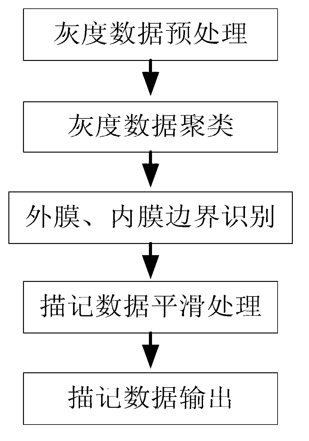
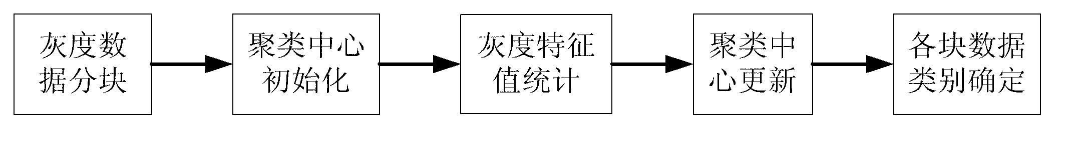
[0056] Such as Figure 1 to Figure 9 As shown, a method for detecting and calculating the boundary of the inner and outer membranes of the vascular tissue structure in the ultrasonic two-dimensional grayscale image is stored in the two-dimensional grayscale data of the blood vessel ultrasound image in the data buffer, and the region of interest is determined according to the buffer area , and follow figure 1 Middle boundary identification and measurement calculation operation, output result display.

[0057] The vascular ultrasound two-dimensional grayscale data involved in the present invention can be the blood vessel grayscale image of any tissue structure of the human body, including but not limited to the two-dimensional ultrasound blood vessel grayscale image of the common carotid artery, internal carotid artery, an...


PUM


Abstract

The invention relates to ultrasound detection and discloses a method of identifying, tracing and measuring external and internal membranes of a vessel. The method of identifying, tracing and measuring the external and internal membranes of the vessel includes the following steps. Firstly, data in a zone of interest are obtained. Thirdly, Gray-scale data clustering is carried out on an image of the zone of interest in order to obtain all target zones. Fourthly, boundaries of the external and internal membranes of the vessel are traced. Sixthly, thickness of the vessel is worked out. The method of identifying, tracing and measuring the external and internal membranes of the vessel has the advantages of improving calibration precision of the external and internal membranes of the vessel and media as well as reliability of calculation on irregular vessel function detecting indexes.

Description

technical field [0001] The invention relates to ultrasonic detection, in particular to a method for identifying, tracing and measuring the intima and intima of blood vessels. Background technique [0002] Vascular atherosclerosis can cause arterial narrowing, that is, coronary heart disease, which can cause shortness of breath, angina pectoris, and even heart disease and other diseases. life property. It is difficult to measure the degree of atherosclerosis, and there are certain risks, but the carotid artery has been proven to effectively reflect the degree of atherosclerosis. [0003] Because the B-mode ultrasonic scanning technology can provide high-resolution images of carotid arteries, the scanning will not cause harm to the human body and the cost is low. Ultrasound images can provide clear multi-layer digital image data of the carotid artery wall. Through the measurement and analysis of this data, the degree of arterial sclerosis can be estimated. Therefore, the acc...

Claims


Application Information

Patent Timeline
Patent Type & Authority Applications(China)
IPC IPC(8): G06K9/00G06K9/62A61B8/08G06V10/44
CPCG06T2207/30101G06T7/12G06V10/44G06F18/23213
Inventor 周传涛马景辉刘秀坚
Owner SHENZHEN EMPEROR ELECTRONICS TECH
Why Eureka
Try Eureka