Method using cross-sectional image to obtain coordinate of target point in stereotaxic apparatus

A stereotaxic instrument and tomographic image technology, which can be used in computer tomography scanners, stereotaxic surgical instruments, diagnosis, etc., can solve problems such as low resolution, error, and large scanning layer thickness.

Inactive Publication Date: 2013-05-08
ZHEJIANG UNIV
View PDF4 Cites 8 Cited by
  • Summary
  • Abstract
  • Description
  • Claims
  • Application Information

AI Technical Summary

Problems solved by technology

However, the effect of reconstruction is affected by the large thickness of the scanning layer (MRI is usually 3 mm), low resolution (the fine structure of the basal ganglia in CT images is not clearly displayed), and the influence of the three-dimensional reconstruction algorithm. The AC-PC plane obtained by reconstruction There is a problem of missing or incorrect information

Method used


Image

  • Method using cross-sectional image to obtain coordinate of target point in stereotaxic apparatus
  • Method using cross-sectional image to obtain coordinate of target point in stereotaxic apparatus
  • Method using cross-sectional image to obtain coordinate of target point in stereotaxic apparatus

Examples


Embodiment Construction

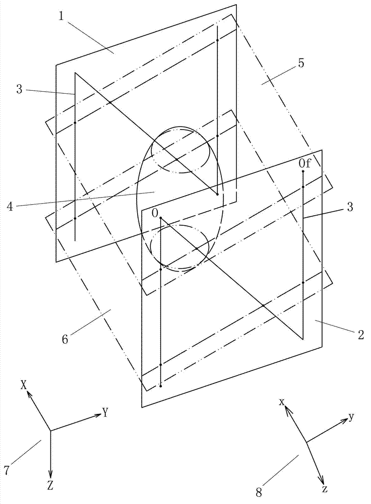
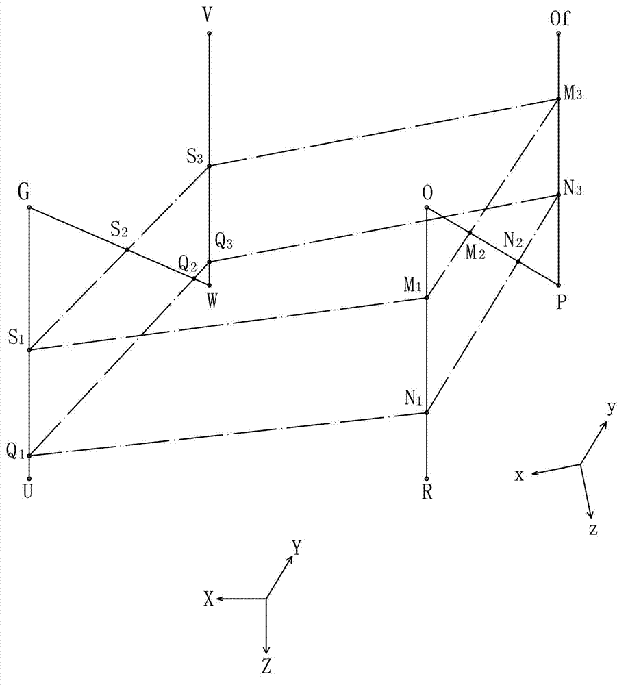
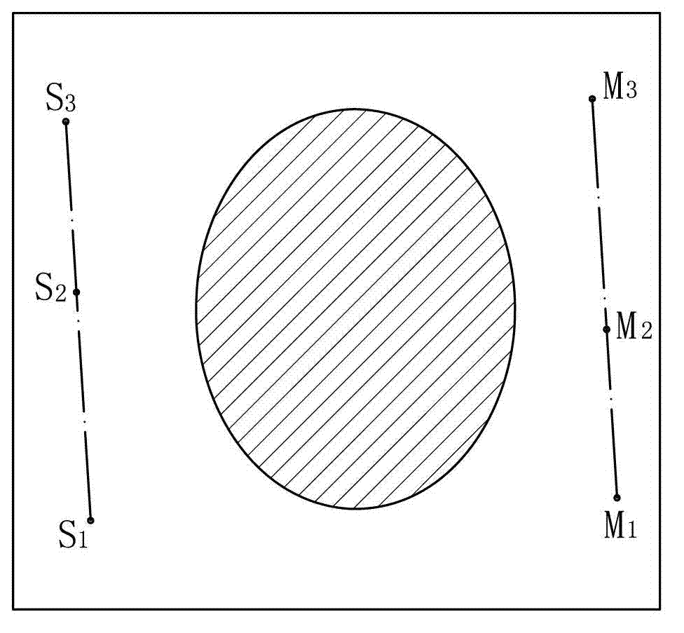
[0018] like figure 1 As shown, the positioning plates 1 and 2 on both sides of the frame of the stereotaxic apparatus have an "N"-shaped metal wire 3 (metal wire for CT positioning, developing materials such as magnesium sulfate for MRI positioning). In the state that the frame is installed, the origin of the frame coordinates is located at the upper right back of the patient's skull 4, which corresponds to the vertex O of the "N" shape wire, and the scale of the orientation instrument also starts from this origin. The coordinate direction of the stereotaxic instrument is shown in Figure 7, the Y axis is forward, the X axis is left, and the Z axis is downward. The fault position during tomographic scanning is shown in Figure 6 and 7. The coordinate direction of the tomographic image is as follows figure 1 As shown in middle 8, on the tomographic image, x is to the left, y is to the front, and z is the downward direction perpendicular to the fault. like figure 2 As shown, t...


PUM


Abstract

The invention provides a method using a cross-sectional image to obtain a coordinate of a target point in a stereotaxic apparatus. The method comprises the steps: (1) selecting the cross-sectional image where six mark points of N-shaped metal lines of two positioning plates can be completely seen in the cross-sectional image, and measuring a coordinate parameter of the mark points; (2) establishing an equation in an image coordinate system of a Y-Z plane, an X-Z plane and an X-Y plane of a stereotaxic apparatus frame; (3) selecting the target point on the cross-sectional image, obtaining the three-dimension coordinate parameter in the image coordinate system, and calculating the distance from the target point to the Y-Z plane, the X-Z plane and the X-Y plane of the frame, and value of the distance is an X, Y and Z coordinate in the stereotaxic apparatus frame coordinate system of the target point. The method reduces installation difficulty of the stereotaxic apparatus frame, reduces difficulty of a stereotaxis operation, and especially is suitable for coordinate positioning of an intercranial 'invisible' target point.

Description

technical field [0001] The invention relates to a method for obtaining the coordinates of a target point in a stereotaxic apparatus by using a tomographic image, which can be applied to the determination of the target point coordinates of a hybrid stereotaxic apparatus, and is especially suitable for Leksell and similar stereotaxic apparatuses. Background technique [0002] The traditional stereotaxic neurosurgery technology uses a mechanical stereotaxic instrument, and the frame of the stereotaxic instrument is fixed on the patient's head during the MRI or CT scan. Through the measurement and calculation of the MRI or CT images, the surgical target is determined. Coordinates to guide the operation. Intracranial lesions that can be displayed on MRI or CT images are called "visible targets", and those that cannot be displayed due to the resolution of the machine are called "invisible targets". [0003] There is an "N"-shaped metal wire (metal wire for CT positioning, magnesi...

Claims


Application Information

Patent Timeline
Patent Type & Authority Applications(China)
IPC IPC(8): A61B19/00A61B5/055A61B6/03A61B90/10
Inventor 张建民刘凤强傅伟明苏英
Owner ZHEJIANG UNIV
Why Eureka
Try Eureka