Detection method for radiosensitivity of solid tumor cell

A detection method and technology of tumor cells, applied in the field of detection of cell radiosensitivity of solid tumors, can solve problems such as complex operation, stability, and low success rate, and achieve the effect of deepening understanding

Inactive Publication Date: 2013-05-08
CANCER INST & HOSPITAL CHINESE ACADEMY OF MEDICAL SCI +1
View PDF1 Cites 5 Cited by
  • Summary
  • Abstract
  • Description
  • Claims
  • Application Information

AI Technical Summary

Problems solved by technology

Due to the complicated operation, low stability and low success rate of these experimental methods, most of them remain at the le

Method used


Image

  • Detection method for radiosensitivity of solid tumor cell
  • Detection method for radiosensitivity of solid tumor cell
  • Detection method for radiosensitivity of solid tumor cell

Examples


Embodiment 1

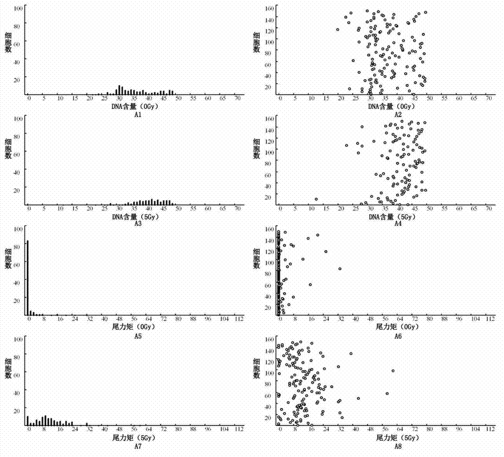
[0048] Figure 1 is a legend of the analysis report of the radiosensitivity of the cells of the nasopharyngeal carcinoma samples. 1A1-A8 are analysis reports of lymphoma cells cultured in vitro as external reference controls, which are used as positive references for system error correction and radiosensitivity assessment. From Figure 1A1 to Figure 1A8, the histogram and scatter diagram of the DNA content distribution of cells under 0 irradiation, the histogram and scatter diagram of the DNA content distribution of cells after 5Gy irradiation, and the distribution of tail moment (TM) of cells under 0 irradiation Histogram and scatter plot and the histogram and scatter plot of the distribution of cell tail moments after 5Gy irradiation. As shown in the figure, under the conditions of this experiment, the DNA content of lymphoma cells is mostly distributed on the right side of lane 35-40. And it can be seen from the TM distribution diagram that the TM value of the 5Gy irradiatio...

Embodiment 2

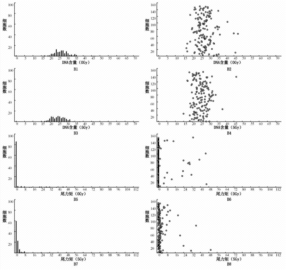
[0052] Figure 2 is a legend of the analysis report showing a typical high radiosensitivity (radiosensitive type) of a nasopharyngeal carcinoma sample after cell radiosensitivity testing. The analysis results showed that the TM values ​​of most cells (including normal / tumor cells) in the tumor sample tissue after 5Gy irradiation were relatively large, even greater than that of the external reference control cells after 5Gy irradiation (shown as obvious under the microscope). "comet tail" formation). Fig. 2A1-A8, B1-B8 and C1-C8 are respectively the analysis reports of normal cells, tumor cells and external reference control lymphoma cells of test samples, as shown in the figure, the TM difference of external reference control lymphoma cells is 21.07, the TM difference of normal cells was 23.6, and the radiosensitivity was 1.12 (23.6 / 21.07), and the TM difference of tumor cells was 42.36, and the radiosensitivity was 2.01 (42.36 / 21.07).

[0053] The actual radioreactivity measu...

Embodiment 3

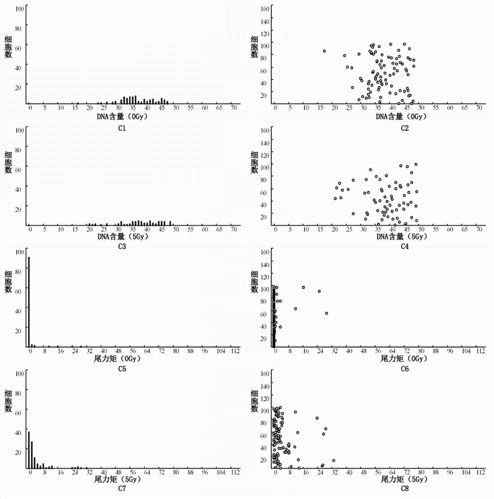
[0055] Figure 3 is a legend of the analysis report showing a typical low radiosensitivity (radiation insensitive type) of a nasopharyngeal carcinoma sample after cell radiosensitivity testing. The analysis results showed that the TM values ​​of normal cells (lymphocytes, fibroblasts) and tumor cells in the tumor sample tissue after 5Gy irradiation were much smaller than those of the external reference control cells after 5Gy irradiation (shown as no obvious under the microscope). "comet tail" formation). Figure 3A1-A8, B1-B8, and C1-C8 are the analysis reports of normal cells, tumor cells, and lymphoma cells of the external reference control, respectively. As shown in the figure, the TM difference of the external reference control lymphoma cells is 73.24, The TM difference of normal cells was 1.13 and the radiosensitivity was 0.02 (1.13 / 73.24), while the TM difference of tumor cells was 13.03 and the sensitivity was 0.18 (13.03 / 73.24).

[0056] The actual radioreactivity meas...


PUM


Abstract

The invention relates to a detection method for radiosensitivity of a solid tumor cell. The detection method comprises the steps of taking detected tumor sample tissues, and preparing the detected tumor sample tissues into single-cell suspensions; taking lymphoma cells subjected to in vitro suspension cultivation as outer contrasts; respectively dividing detected tumor samples and the single-cell suspensions of the lymphoma cells taken as the outer contrasts into an illumination group and a blank control group, and carrying out single-pass illumination on the single-cell suspensions in the illumination group by adopting an X ray in a dosage of 5 Gy, wherein the single-cell suspensions in the blank control group are not subjected to radioactive ray illumination; carrying out cell disruption and rinsing on each group of the single-cell suspensions; carrying out electrophoresis on each group of the cells after disruption; carrying out PI (Propidium Iodide) dyeing on each group of the cells after electrophoresis; under a fluorescence microscope, respectively carrying out tumor/normal cell classification image capturing on the cells in the illumination group and the blank control group of the detected tumor samples, and carrying out image capturing on outer contrast cells; and carrying out data processing on images of the tumor cells, the normal cells and the outer contrast cells in the cells of the detected tumor samples, thus judging the radiosensitivity of the cells of the detected tumor samples.

Description

technical field [0001] The present invention relates to a method for detecting the radiosensitivity of solid tumor cells. Background technique [0002] The radiosensitivity detection of solid tumor cells is the basis for the study and prediction of the radiosensitivity of solid tumor cells. The prerequisite is to establish a sensitive, stable and operable radiosensitivity detection method for solid tumor cells. For this reason, several major radiobiological research centers abroad have successively carried out studies on predicting tumor radiosensitivity over the years, and have introduced some potentially valuable detection methods, such as SF2 (cell survival fraction after 2Gy irradiation), Tpot ( Potential doubling time), micronucleus rate, primary cell clone formation analysis and cell adhesion analysis, etc. Due to the complicated operation, low stability and low success rate of these experimental methods, so far most of them remain at the level of cultured cells in vi...

Claims


Application Information

Patent Timeline
IPC IPC(8): G01N23/00G01N21/64
Inventor 高黎杨伟志房超
Owner CANCER INST & HOSPITAL CHINESE ACADEMY OF MEDICAL SCI
Why Eureka
Try Eureka