
Set of glass slides used for carrying out immunohistochemistry staining and storage to tissue slice

A technique of tissue slicing and immunochemistry, which is applied in the field of medical biological experiment equipment, can solve the problems of slice falling off, cover glass shifting, tissue transfer damage, etc., and achieve the effect of ensuring stability

Inactive Publication Date: 2013-05-08
THE THIRD AFFILIATED HOSPITAL OF THIRD MILITARY MEDICAL UNIV OF PLA
View PDF8 Cites 14 Cited by
  • Summary
  • Abstract
  • Description
  • Claims
  • Application Information

AI Technical Summary

Problems solved by technology

[0003] The technical problem to be solved by the present invention is to propose a set of slides that can be used in the process of immunochemical staining of tissues, which may easily cause slices to fall off, antibody overflow, cover glass shift, and tissue transfer damage. Paraffin or frozen tissue immunochemical staining and effectively preserved glass slides, and the size of tissue cells can be initially measured through the matching sealing unit

Method used


Image

  • Set of glass slides used for carrying out immunohistochemistry staining and storage to tissue slice
  • Set of glass slides used for carrying out immunohistochemistry staining and storage to tissue slice

Examples


Embodiment Construction

[0014] The present invention will be described in detail below in conjunction with the accompanying drawings and specific examples.

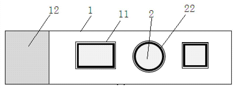
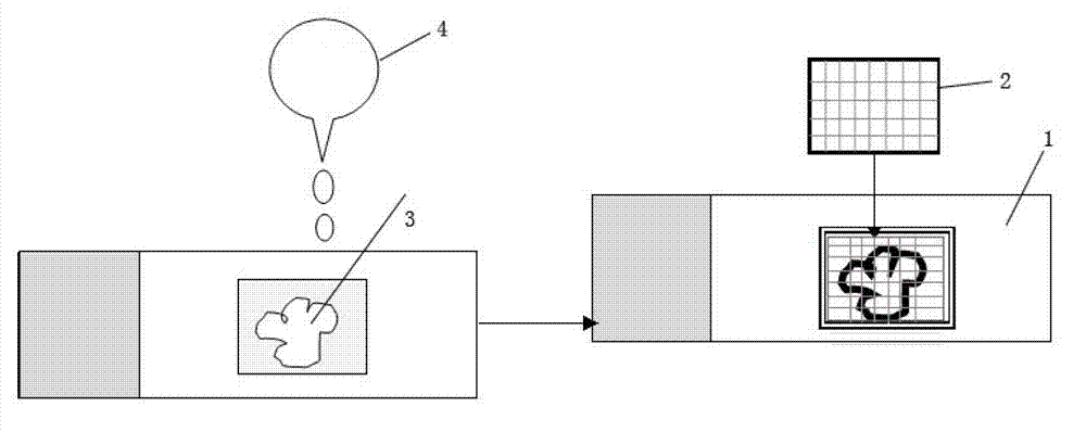
[0015] Such as figure 1 and figure 2 As shown, the slide glass is composed of an object loading unit 1 and a sealing unit 2 . The loading unit 1 is glass with a thickness of 1.2mm. It has grooves 11 of different depths and shapes (such as circle, ellipse, rectangle and square, etc.) on its bearing surface. The depth of the groove is 600um-800um, and the inner surface is smooth. For accommodating tissue slices, each groove corresponds to a cover-slice unit 2 whose shape and depth can match it, and the thickness of the cover-slice unit is 200 um. On the sealing unit, with its center as the base point, vertical and horizontal vertical intersecting straight lines are distributed, dividing a plurality of squares 21 with a side length of 10 um, and a sealing ring 22 is provided on the edge of the sealing unit. On the left side of the loading surfa...


PUM


Abstract

Disclosed is a set of glass slides used for carrying out immunohistochemistry staining and storage to a tissue slice. A bearing face of an object bearing unit is provided with grooves which are different in depth and shape and are used for containing the tissue slice. The inner surfaces of the grooves are smooth. Each groove is corresponding to a mounting unit. A mounting unit sheet is matched with the shapes of the grooves. Straight lines which are vertically crossed in a vertical and horizontal mode use the centers of the mounting units as a base point and are arranged on the mounting units, each square is formed, the side length of each square is 10 micrometers, and sealing rings are arranged on the edges of the mounting units. The set of glass slides effectively avoids the problems that a tissue slice is easy to drop off, an antibody overflows, cover glass shifts, and tissue transfer is damaged in an existing immunohistochemistry detection process, is simple in structure, low in cost, and capable of well ensuring stability of the tissue slice and the cover glass and effectiveness of contact reaction of the antibody and a tissue, and can measure and calculate the size and the area of a detected tissue through scale marks on the mounting units.

Description

technical field [0001] The invention relates to a medical biological experiment device, in particular to a set of glass slides for performing immunohistochemical staining on tissue sections. technical background [0002] In medical experiments, using the principle of specific binding of antigens and antibodies, immunochemical staining of tissues with different antibodies is one of the most commonly used methods to detect functional changes in the body under physiological or pathological conditions. During the immunostaining process of tissue sections, it is usually stained by patch method and bleach method. Staining by the patch method firstly slices paraffin tissue or frozen tissue, pastes it on a glass slide, and then detects it according to the relevant steps of immunohistochemical staining. At present, the detection method usually has the following problems: 1. Repeated washing of the tissue during the detection process may easily cause tissue slices to fall off; 2. Sin...

Claims


Application Information

Patent Timeline
Patent Type & Authority Applications(China)
IPC IPC(8): G02B21/34G01N1/31
Inventor 刘媛叶有义谭波涛王莉伍亚民王正国
Owner THE THIRD AFFILIATED HOSPITAL OF THIRD MILITARY MEDICAL UNIV OF PLA
Learn More