Three-dimensional ultrasonic positioning and surgery navigation system as well as image processing method thereof

A technology of surgical navigation and three-dimensional ultrasound, which is applied in the field of image processing and ultrasound image processing system to achieve the effect of automation

Inactive Publication Date: 2013-05-15
JIANGSU YADA TECH GRP
View PDF0 Cites 20 Cited by
  • Summary
  • Abstract
  • Description
  • Claims
  • Application Information

AI Technical Summary

Problems solved by technology

[0005] In terms of surgical treatment, the Mammotome surgery in the United States is relatively advanced, but this technology only involves surgery, and the conventional B-ultrasound positioning method is still used

Method used


Image

  • Three-dimensional ultrasonic positioning and surgery navigation system as well as image processing method thereof

Examples


Embodiment Construction

[0017] The technical solution of the present invention will be described in detail below through a best embodiment in conjunction with the accompanying drawings, but the protection scope of the present invention is not limited to the embodiment.

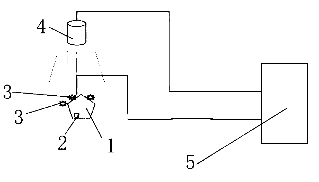
[0018] Such as figure 1 As shown, a three-dimensional ultrasonic positioning and surgical navigation system includes an ultrasonic probe 1, an infrared probe 2 is arranged on the ultrasonic probe 1, and the infrared probe 2 is equipped with three non-collinear infrared beads 3, and also includes infrared The positioning device 4 is used to determine the position of the infrared ball to locate the infrared probe. The ultrasonic probe, the infrared probe and the infrared positioning device are connected to the computer 5, and the ultrasonic probe is a B-ultrasound probe.

[0019] The image processing method of the three-dimensional ultrasonic positioning and surgical navigation system, the construction and update of data, the infrared posit...


PUM


Abstract

The invention discloses a three-dimensional ultrasonic positioning and surgery navigation system. The three-dimensional ultrasonic positioning and surgery navigation system comprises an ultrasonic probe, wherein an infrared probe is arranged on the ultrasonic probe; and non-collinear three small infrared balls are mounted on the infrared probe. The three-dimensional ultrasonic positioning and surgery navigation system further comprises an infrared positioning device for positioning the infrared probe by measuring the positions of the small infrared balls; and the ultrasonic probe, the infrared probe and the infrared positioning device are connected with a computer. The invention further discloses an image processing method of the three-dimensional ultrasonic positioning and surgery navigation system. The image processing method can be used for virtually displaying three-dimensional scenes, such as a breast, a breast tumor, a puncture needle, and a surgery path, through data establishment and update, elastic registration and virtual reality. The three-dimensional ultrasonic positioning and surgery navigation system provided by the invention carries out data treatment after collecting data through ultrasonic waves and the infrared probe, is used for accurately positioning the breast by using the ultrasonic waves and infrared positioning, is used for diagnosing breast diseases, and further can be used for navigating breast puncture and surgery.

Description

Technical field [0001] The invention relates to a medical device, in particular to an ultrasonic image processing system and an image processing method of the system. Background technique [0002] At present, there are several examination methods for breast tumors in China: ① physical examination, ② mammogram X-ray, ③ ultrasound, ④ infrared scan, ⑤ puncture, ⑥ biopsy, ⑦ Others: including CT, MRI. In the past, the diagnosis of breast tumors mainly relied on X-ray photography. X-ray mammography can find occult lesions including occult breast cancer, and can perform stereotactic needle biopsy. However, the mammary glands of middle-aged and young women are mostly dense, and the contrast between fibroadenoma and surrounding tissues is not great. Therefore, mammography is not the preferred method for detecting benign breast tumors. Compared with mammography X-ray examination, ultrasound is not affected by tissue density, especially when dense breasts are accompanied by tumors, the con...

Claims


Application Information

Patent Timeline
Patent Type & Authority Applications(China)
IPC IPC(8): A61B19/00A61B34/20
Inventor 刘永君刘凯葛云章东易际平何侠翟振宇
Owner JIANGSU YADA TECH GRP
Why Eureka
Try Eureka