Histological classification immunohistochemical multiple staining detection method for lung cancer

A detection method and immunohistochemical technology, applied in the field of lung cancer histological typing immunohistochemical multiple staining detection, can solve the problems of difficult typing, time-consuming and labor-intensive detection of immunohistochemical techniques separately, and achieve less staining steps and better experimental results Intuitive contrast, high stability effect

Active Publication Date: 2013-06-12
FUZHOU MAIXIN BIOTECH CO LTD
View PDF2 Cites 13 Cited by
  • Summary
  • Abstract
  • Description
  • Claims
  • Application Information

AI Technical Summary

Problems solved by technology

[0005] The purpose of the present invention is to overcome the disadvantages of difficult typing of lung cancer histology and the time-consuming and laborious det

Method used


Image

  • Histological classification immunohistochemical multiple staining detection method for lung cancer
  • Histological classification immunohistochemical multiple staining detection method for lung cancer
  • Histological classification immunohistochemical multiple staining detection method for lung cancer

Examples


Embodiment 1

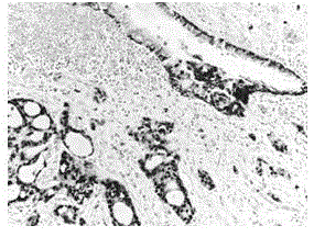
[0027] The research object was formaldehyde-fixed-paraffin-embedded lung adenocarcinoma tissue (Department of Pathology, Fujian Provincial Hospital). The immunohistochemistry experiment procedure is as follows:

[0028] (1) Place the tissue slices in a 67°C incubator for 2 hours.

[0029] (2) Routine xylene dewaxing 3 times, 6 minutes each time, hydration in 100%, 100%, 95%, 85% gradient ethanol, 3 minutes each time, and finally rinse with tap water.

[0030] (3) Boil directly in pH9.0 EDTA antigen retrieval solution, cool to room temperature naturally, rinse with tap water and rinse with PBS for 3×3 minutes.

[0031] (4) Incubate in 3% hydrogen peroxide for 10 minutes at room temperature to block endogenous peroxidase, and rinse with PBS for 3×3 minutes.

[0032] (5) Block with normal animal serum for 10 minutes, discard the excess serum, without washing, add Napsin A, TTF-1, Desmoglein 3 mixed primary antibody dropwise, and the antibody titers in the mixed primary antibody are Napsin...

Embodiment 2

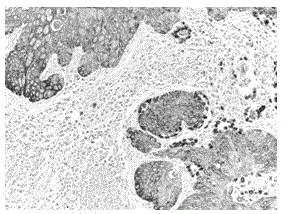
[0039] The object of study was formaldehyde-fixed-paraffin-embedded lung squamous cell carcinoma tissue (Department of Pathology, Fujian Provincial Hospital). The immunohistochemistry experiment procedure is as follows:

[0040] (1) Place the tissue slices in a 67°C incubator for 2 hours.

[0041] (2) Routine xylene dewaxing 3 times, 6 minutes each time, hydration in 100%, 100%, 95%, 85% gradient ethanol, 3 minutes each time, and finally rinse with tap water.

[0042] (3) Boil directly in pH9.0 EDTA antigen retrieval solution, cool to room temperature naturally, rinse with tap water and rinse with PBS for 3×3 minutes.

[0043] (4) Incubate in 3% hydrogen peroxide for 10 minutes at room temperature to block endogenous peroxidase, and rinse with PBS for 3×3 minutes.

[0044] (5) Block with normal animal serum for 10 minutes, discard the excess serum, without washing, add Napsin A, TTF-1, CK5 / 6 mixed primary antibody dropwise, and the antibody titers in the mixed primary antibody are Naps...

Embodiment 3

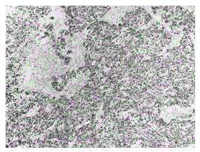
[0051] The object of study was formaldehyde-fixed-paraffin-embedded lung small cell carcinoma (Department of Pathology, Fujian Provincial Hospital). The immunohistochemistry experiment procedure is as follows:

[0052] (1) Place the tissue slices in a 67°C incubator for 2 hours.

[0053] (2) Routine xylene dewaxing 3 times, 6 minutes each time, hydration in 100%, 100%, 95%, 85% gradient ethanol, 3 minutes each time, and finally rinse with tap water.

[0054] (3) Boil directly in pH9.0 EDTA antigen retrieval solution, cool to room temperature naturally, rinse with tap water and rinse with PBS for 3×3 minutes.

[0055] (4) Incubate in 3% hydrogen peroxide for 10 minutes at room temperature to block endogenous peroxidase, and rinse with PBS for 3×3 minutes.

[0056] (5) Block with normal animal serum for 10 minutes, discard excess serum, without washing, add Napsin A, TTF-1, p40, TRIM29 mixed primary antibody dropwise, and the antibody titers in the mixed primary antibody are Napsin A 1:2...


PUM

PropertyMeasurementUnit
Thicknessaaaaaaaaaa

Abstract

The invention belongs to the technical field of immunohistochemistry, and in particular relates to a histological classification immunohistochemical multiple staining detection method for lung cancer. The primary antibody in the detection method is various mixed primary antibodies. In order to overcome the defects that existing lunge cancer is not easy to classify in histology, and detection based on an immunohistochemical technology consumes time and wastes labor, the staining process is simple and quick by adopting the method, so that more abundant information with strong contrast can be obtained on a same slice quickly and conveniently. According to the invention, the mixed primary antibodies and various detection amplifying systems are applied to identifying histological classification of lung cancer, so that not only can the same sensitivity and specificity of each antibody in the mixed antibodies be maintained in comparison with those in simple staining, but also the method has the advantages of less staining steps, short experimental time, high stability, intuitionistic comparison of experimental result, far great information amount than single staining and the like, especially has remarkable advantages of biopsic pathological diagnosis with small specimen amount.

Description

Technical field [0001] The present invention belongs to the technical field of immunohistochemistry. Specifically, the present invention relates to an immunohistochemical multiple staining detection method for histological classification of lung cancer. Background technique [0002] Lung cancer is the most common malignant tumor of the respiratory system and the most common cancer that causes death. It can be divided into small cell lung cancer and non-small cell lung cancer. The common histological types of the latter are squamous cell carcinoma, adenocarcinoma and large cell carcinoma, and occasionally some other types or mixed cell types with a lower incidence. The distinction between small cell lung cancer and non-small cell carcinoma has always been regarded as the most important problem in pathological diagnosis. Distinguishing between the two has important therapeutic significance. The pathologist makes the differential diagnosis of the two based on cell morphology and imm...

Claims


Application Information

Patent Timeline
IPC IPC(8): G01N33/574
Inventor 杨清海王小亚
Owner FUZHOU MAIXIN BIOTECH CO LTD
Why Eureka
Try Eureka