Device and method for structure-reconstruction-based subcutaneous vein three-dimensional visualization

A subcutaneous vein, three-dimensional technology, applied in the medical field, can solve the problems of dependence, difficulty in measuring the relationship between the three-dimensional structure of veins and blood vessels and the relative position of the skin, and the inability to provide blood vessel depth information, so as to achieve the effect of ensuring accurate extraction

Active Publication Date: 2013-10-02
BEIJING INSTITUTE OF TECHNOLOGYGY
View PDF4 Cites 75 Cited by
  • Summary
  • Abstract
  • Description
  • Claims
  • Application Information

AI Technical Summary

Problems solved by technology

[0008] 1. The method of two-dimensional screen display or two-dimensional projection display can only perceive the two-dimensional distribution characteristics of veins and blood vessels, and cannot provide depth information of blood vessels
[0009] 2. The vein display based on two-dimensional imaging technology is very dependent on the imaging angle, and

Method used


Image

  • Device and method for structure-reconstruction-based subcutaneous vein three-dimensional visualization
  • Device and method for structure-reconstruction-based subcutaneous vein three-dimensional visualization
  • Device and method for structure-reconstruction-based subcutaneous vein three-dimensional visualization

Examples


Embodiment Construction

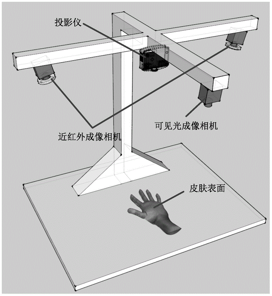
[0034] The result figure of the hardware image acquisition system of the present invention is as attached figure 1 As shown, it includes 1 industrial camera for visible light imaging, 2 industrial cameras equipped with near-infrared light sources and infrared filters, 1 short-distance projector and a bracket for placing the above devices.

[0035]Projector and visible light imaging camera constitute the hardware equipment for 3D reconstruction of skin. The projector is responsible for projecting the computer-coded structured light image; the industrial camera is responsible for collecting the structured light image projected on the skin surface and modulated by the skin. There is a fixed positional relationship between the projector and the industrial camera, and they are fixed together on the same rigid crossbar, which ensures the uniqueness of the 3D reconstruction process. During the calibration process, it is necessary to adjust the focal length and installation angle so ...


PUM

PropertyMeasurementUnit
Peak wavelengthaaaaaaaaaa

Abstract

The invention discloses a device and a method for structure-reconstruction-based subcutaneous vein three-dimensional visualization, which can acquire three-dimensional space information of a vein and skin. The method comprises the following steps: step 1, performing parameter calibration for three cameras; step 2, utilizing calibrated cameras to perform parameter calibration for the projector; step 3, utilizing structured light method to realize three-dimensional reconstruction of skin surface; step 4, adopting a template matching method to extract a vein central line in near-infrared images; step 5, adopting a pole line matching method to realize three-dimensional reconstruction of the vein central line; step 6, adopting a light projected method accelerated by a GPU (Graphics Processing Unit) to realize three-dimensional visualization of an opisthenar and the vein on a computer display; finally, simulating to obtain a projected image with the projector as a viewpoint according to the three-dimensional data, and projecting the image to the skin surface to be inspected to realize two-dimensional projected display.

Description

technical field [0001] The present invention relates to a venous blood vessel display device, in particular to a 3D visualization device based on 3D reconstruction data of vein and skin surface and a 2D projected blood vessel enhanced display device, which are mainly used in medical fields such as subcutaneous intravenous injection and vein structure teaching. Background technique [0002] In the medical field, with the continuous development of imaging technology, the limitations of visible light imaging perceived by the human eye are increasingly exposed, and a large amount of information of the detected tissue is hidden in the invisible spectral band. Therefore, imaging technologies based on infrared, ultraviolet, and X-ray bands are more and more widely used in the field of tissue detection and identification due to their superior imaging characteristics. When applied to subcutaneous vein extraction, compared with the low contrast of vein images under visible light imagi...

Claims


Application Information

Patent Timeline
IPC IPC(8): G06T7/00G06T17/00A61B5/00
Inventor 杨健王涌天刘越宋宪政
Owner BEIJING INSTITUTE OF TECHNOLOGYGY
Why Eureka
Try Eureka